Resource Libraries
Ehlers-Danlos Syndrome Dental Problems: What to Know
How Ehlers-Danlos syndrome affects the mouth, from TMJ instability and fragile gums to bleeding and local anesthetic failure, and how to plan safer care.
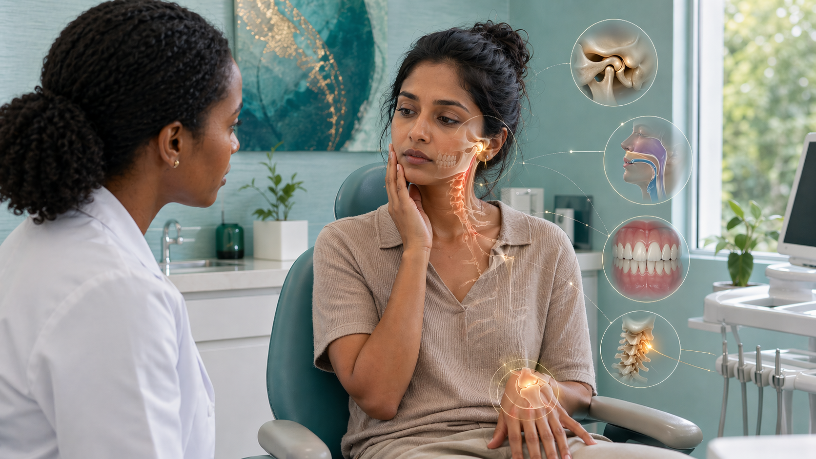
Hypermobility, Ehlers-Danlos Syndrome, and Dentistry: What Patients and Clinicians Need to Know
For many people with hypermobility or Ehlers-Danlos syndrome, dental care does not feel routine. A cleaning may leave the jaw sore for days. The mouth may not open comfortably for very long. Local anesthetic may not work the way it should. Tissues may bruise, tear, or heal differently. Teeth may crowd, shift, or relapse after orthodontic treatment. These are not random complaints. They may all trace back to one bigger issue: connective tissue affects far more than just the knees, shoulders, and elbows. It also affects the mouth, the jaw joints, the gums, the airway, and how the body tolerates dental treatment.
That is why this topic matters. Ehlers-Danlos syndromes, or EDS, are a group of inherited connective-tissue disorders. Hypermobility spectrum disorders, or HSD, are related conditions that can also come with real symptoms and real mechanical problems. In the mouth, these conditions can affect jaw stability, oral tissues, tooth support, healing, pain, bleeding, and sleep-related breathing. The mistake dentistry often makes is treating these patients like everyone else and then acting surprised when routine treatment does not go routinely.
First, not all EDS is the same
This is one of the most important points in the whole discussion. People often say “EDS” as though it is one condition with one oral pattern. It is not. Hypermobile EDS is different from periodontal EDS, and that distinction matters a lot in dentistry. GeneReviews notes that people with hypermobile EDS commonly have dental crowding, a high and narrow palate, TMJ dysfunction, and frequent reports that local anesthetics do not work well. But it also states that severe periodontal disease and early tooth decay not explained by lifestyle are not common in hEDS and should raise concern for periodontal EDS instead.
Periodontal EDS is much more centered in the mouth. GeneReviews describes it as a condition where periodontal breakdown can begin in the teen years, with lack of attached gingiva, thin fragile gums, recession, premature tooth loss, and a high risk of peri-implantitis and implant failure. That means a patient with dramatic recession, tooth mobility, or early tooth loss should not automatically be lumped into a vague “hypermobility” box. The subtype matters because the risks are not the same.
Oral and Dental Manifestations (General)
Kapferer-Seebacher I, Schnabl D, Zschocke J, Pope FM
PMID: 32147746
This systematic review cataloged dental findings across EDS subtypes. Classical EDS showed pulp calcification and root shortening. Vascular EDS showed unusual pulp shapes, extra-long roots, and fused molar roots.
Lepperdinger U, Zschocke J, Kapferer-Seebacher I
PMID: 34741498
Summarized oral problems across EDS types including periodontitis differences between subtypes, soft tissue fragility, dental anomalies, and implant concerns. Emphasized the need for genetic confirmation and specialized dental care.
Grand A, Fournier BPJ, Chevalier G, Benistan K, Ferré FC
PMID: 41840838
Compared 14 genetically confirmed EDS patients to 30 controls. EDS patients had significantly more TMJ disorders, gingivitis, tongue habits, frenula atrophy, pulp changes, and root shortening than healthy people.
David A, Romain N, Marine B, Joël F
PMID: 40615270
A systematic review of oral/facial features across all EDS subtypes. Found subtype-specific patterns including tooth agenesis, root fusion, pulp calcifications, and gingival overgrowth, showing that a dental exam can help identify which type of EDS someone has.
Mitakides J, Tinkle BT
PMID: 28192626
Reviewed how EDS affects the mouth and jaw, including soft tissue fragility, TMJ dysfunction, dental crowding, and the connection between jaw problems and whole-body musculoskeletal issues. Highlighted that these features are often overlooked.
Yekkalam N, Sipilä K, Novo M, et al.
PMID: 39352367
Surveyed 133 women with confirmed hEDS/HSD and TMJ symptoms. Found that jaw pain, clicking, and grinding significantly hurt their oral quality of life. Poor general health and multiple symptoms made everything worse.
The jaw joint is often part of the problem
One of the clearest ways hypermobility shows up in dentistry is through the temporomandibular joint, or TMJ. People with EDS often report clicking, popping, locking, pain, limited opening, and soreness after long dental visits. In many of these patients, the jaw is not just painful. It is less stable.
That matters because the body may already be compensating before the dental appointment even starts. The muscles of the jaw and neck may be working overtime to stabilize a loose joint. So when that patient is asked to stay open for a long appointment, use a bite block, tolerate strong retraction, or keep the neck in a strained position, the procedure may be much harder on the body than the dentist realizes. The restoration may look beautiful while the patient leaves feeling wrecked. That does not mean the patient is dramatic. It means the body paid a higher mechanical price for the appointment.
There is also growing interest in whether regenerative or stabilizing injections may help some TMJ patients with hypermobility or recurrent dislocation. This is where the conversation needs nuance. The strongest evidence is not for PRF specifically in EDS across all joints. Instead, the current literature suggests three related but distinct things: PRF appears promising for painful TMJ disorders in the general population, autologous blood injection has some of the best evidence for recurrent TMJ dislocation, and prolotherapy has some support for painful TMJ dysfunction and joint hypermobility. In hypermobile EDS itself, GeneReviews mentions prolotherapy as having been shown safe and effective in one study, but that is still a way from saying PRF is a definitive established standard treatment for TMJ instability in EDS.
So the honest takeaway is this: regenerative injections are an interesting and emerging area, especially for patients whose joint instability keeps driving pain or recurrent dislocation, but the EDS-specific scientific evidence is still early. That means the idea is worth discussing, but it should be presented as promising and biologically plausible, not settled. That said, I would be lying if I said this area does not genuinely excite me. PRF is made from the patient’s own blood, which makes it especially appealing from a biological and more holistic perspective. And based on the benefits I have consistently seen from PRF in biological surgeries, I am personally very interested in where this research may go next.
TMJ and Temporomandibular Disorders
Oelerich O, Daume L, Yekkalam N, Hanisch M, Menne MC
PMID: 38603608
This review looked at the connection between Ehlers-Danlos syndrome and jaw joint problems. It found that TMJ disorders like pain, clicking, and disc issues are much more common in people with EDS, especially the hypermobile type.
Bech K, Fogh FM, Lauridsen EF, Sonnesen L
PMID: 35694904
Researchers compared 26 people with hypermobile EDS to 39 healthy controls. The EDS group had significantly more jaw pain, muscle soreness, disc problems, and bone changes in the jaw joints, plus smaller mouth opening.
Willich L, Bohner L, Köppe J, et al.
PMID: 37726791
In 259 EDS patients, about 85% had jaw muscle pain and 46% had a diagnosed TMJ disorder. Having TMJ problems raised the risk of chronic pain 2.5-fold. Over half had critical anxiety scores.
Regenerative Injections for TMJ Hypermobility and Dysfunction
Farshidfar N, Amiri MA, Estrin NE, et al.
PMID: 41006956
This review looked at 23 clinical studies on injectable PRF for TMJ disorders. PRF reduced pain and improved mouth opening, especially after arthrocentesis. It often matched or outperformed PRP and hyaluronic acid, but the authors said better standardized trials are still needed.
de Araújo JSG, de Oliveira da Rosa WL, Meirelles LAD, Dos Santos MG, Boscato N
PMID: 40914979
This 2026 systematic review and meta-analysis found that injectable PRF improved pain and mouth opening in TMJ disorders, especially when combined with arthrocentesis instead of arthrocentesis alone.
Chaulagain RS, Simre SS, Shukla S, et al.
PMID: 40058764
In this randomized clinical trial, arthrocentesis plus PRF worked better than arthrocentesis alone or PRF alone for reducing pain, improving mouth opening, and improving quality of life in TMJ internal derangement.
Nasef M, Alashmawy M, Abdelrahman A, et al.
PMID: 39043507
Patients with TMJ disc displacement without reduction had better pain relief and better mouth opening when PRF was added after arthrocentesis. MRI showed symptom improvement without disc repositioning.
Manafikhi M, Ataya J, Heshmeh O
PMID: 35568935
In this early study, two PRF injections stopped TMJ clicking in all 20 patients by the second injection. Only 2 had recurrence by 6 months. Promising, but still preliminary.
Chęciński M, Chęcińska K, Rąpalska I, et al.
PMID: 37685657
This systematic review found that autologous blood injection prevented further recurrent TMJ dislocations in 75% to 94% of patients. This is one of the strongest evidence areas for TMJ hypermobility-type instability.
Abrahamsson H, Eriksson L, Abrahamsson P, Häggman-Henrikson B
PMID: 31729577
This review concluded that autologous blood injection into the TMJ currently has the best scientific support for recurrent TMJ dislocation.
Sit RW, Reeves KD, Zhong CC, et al.
PMID: 34282199
This meta-analysis found that dextrose prolotherapy worked better than placebo injections for TMJ pain at 12 weeks. Evidence quality was low to moderate, but it supports prolotherapy as a real therapeutic option.
Assiri K, Alqarni A, Almubarak H, et al.
PMID: 40364487
This 2025 clinical study included TMJ dysfunction patients with joint hypermobility. Pain improved and mouth opening increased by 3 months, and the authors recommended prolotherapy for chronic recurrent subluxation and dislocation.
Hakim A — GeneReviews: Hypermobile Ehlers-Danlos Syndrome
PMID: N/A (NBK1279)
GeneReviews notes that prolotherapy, which uses injections to promote scar formation and stability around joints, has been shown safe and effective in one study in hypermobile EDS. That is the strongest direct EDS-specific support in this area.
Turosz N, Chęcińska K, Chęciński M, et al.
PMID: 38792396
This overview of 42 systematic reviews helps organize the hierarchy of evidence: autologous blood appears strongest for recurrent TMJ dislocation, while PRF after arthrocentesis performs better than arthrocentesis alone for painful hypomobility.
Airway issues may be part of the same picture
This is one of the biggest blind spots. Many people think of hypermobility as a joint problem, but it may also affect the tissues that help support the upper airway. GeneReviews for hEDS notes upper-airway and throat issues, and it specifically mentions that TMJ and oral problems may even interfere with use of positive-pressure therapies for sleep apnea.
There is also good evidence that sleep-disordered breathing is more common in people with EDS and generalized joint hypermobility than many clinicians realize. A 2019 meta-analysis found a combined obstructive sleep apnea prevalence of about 48.9% across EDS and joint hypermobility populations.
Why does that matter in dentistry? Because airway problems do not stay neatly in the sleep world. They can show up as mouth breathing, clenching, restless sleep, fatigue, headaches, pain sensitivity, crowded teeth, narrow arches, and poor orthodontic stability. And the airway issue may not always be just “small jaws.” A 2021 cephalometric study found that craniofacial form in hEDS was broadly similar to controls, though airway minimum cross-sectional area and volume tended to be smaller, suggesting that the problem may involve both anatomy and tissue behavior. In other words, some patients may not just have a smaller airway. They may have a more collapsible one.
Sleep Apnea and Airway
Sedky K, Gaisl T, Bennett DS
PMID: 30736885
This meta-analysis combined data from multiple studies and found that about 48.9% of people with EDS or Marfan syndrome had obstructive sleep apnea — roughly six times higher than the general population.
Gaisl T, Giunta C, Bratton DJ, et al.
PMID: 28073822
In 100 EDS patients matched to 100 controls, sleep apnea prevalence was 32% vs. 6%. Craniofacial shape looked similar between groups, suggesting the airway problem may involve tissue behavior, not just bone structure.
Guilleminault C, Primeau M, Chiu HY, et al.
PMID: 23929538
Every EDS patient studied had some form of sleep-disordered breathing on a sleep test. The authors proposed EDS as a genetic model for sleep apnea because collagen defects affect the facial structures that keep the airway open.
Orthodontic relapse may make more sense than many people realize
Some patients with hypermobility feel frustrated that their teeth shift back after braces or aligners, even when they tried hard to follow instructions. This is an area where the science is thinner than many people assume, so it should be discussed honestly. There are case reports and clinical papers describing retention difficulty and special orthodontic considerations in EDS, but we do not have huge modern datasets proving a single relapse pattern across all subtypes.
Still, the concern is biologically reasonable. Teeth do not exist in isolation. They are held in bone by the periodontal ligament and are constantly influenced by the tongue, lips, cheeks, airway, swallowing pattern, and oral posture. If connective tissues are looser, if the airway remains compromised, or if tongue posture and mouth breathing were never addressed, then the end of orthodontic treatment does not automatically mean long-term stability. A 2020 case report described retention difficulty in an adolescent with EDS after treatment, while older papers noted that tooth movement may proceed normally but stability and tissue tolerance can still be problematic.
That means relapse should not always be dismissed as simple noncompliance. Sometimes the teeth were moved, but the body they live in is still pulling in the old direction. For patients, that can feel discouraging. For clinicians, it should mean more honest conversations about retention, airway, oral posture, and the fact that these cases may need more long-term support.
Orthodontic Relapse and Retention in EDS
Gupta S, Ardeshna A, Rossouw PE, Valiathan M
PMID: 39244251
This textbook chapter reviewed systemic conditions that affect orthodontic outcomes. It specifically states that Ehlers-Danlos syndrome poses challenges related to collagen fragility, rapid tooth movement, and orthodontic relapse.
Norton LA
PMID: 6590606
One of the earliest case reports of orthodontic treatment in an EDS patient. Described how tooth movement proceeded but noted special clinical considerations related to tissue response, including the expected behavior of the periodontal ligament in defective collagen.
Norton LA, Assael LA
PMID: 9009927
Described orthodontic and TMJ considerations in EDS patients with short clinical case reports. Noted that while teeth can be moved orthodontically, the connective tissue abnormalities create challenges for stability and TMJ management during and after treatment.
Ganburged G, Suda N, Saito M, et al.
PMID: 20714769
Studied mice with defective fibrillin-1 (a connective tissue protein related to Marfan syndrome). Found disorganized collagen fibers and dilated blood vessels in the periodontal ligament, showing how connective tissue defects directly change the structures that hold teeth in place.
Lin JD, Ryder M, Kang M, Ho SP
PMID: 31850635
Reviewed how the periodontal ligament adapts to mechanical forces. Explains that when connective tissue properties change, the stiffness gradients at the ligament-bone interface shift, which can alter how teeth respond to and recover from orthodontic forces.
DeLeonibus A, Bogart J, Gharb BB, Rampazzo A
PMID: 36683425
Case report and literature review of orthognathic surgery in hEDS. Noted that EDS patients are historically considered poor surgical candidates due to delayed healing and tissue fragility, but showed that jaw surgery can succeed with careful planning. Relevant because orthodontic-surgical cases in EDS carry additional stability concerns.
EDS, Mouth Breathing, and Sleep-Disordered Breathing
Guilleminault C, Primeau M, Chiu HY, et al.
PMID: 23929538
Every EDS patient studied had sleep-disordered breathing on a sleep test. Proposed EDS as a genetic model for obstructive sleep apnea because collagen defects affect the nasal and facial cartilages that shape the airway, promoting mouth breathing and airway collapse.
Sedky K, Gaisl T, Bennett DS
PMID: 30736885
Meta-analysis finding about 48.9% OSA prevalence across EDS and Marfan populations — roughly six times higher than the general population. The authors attributed this to both bony and soft-tissue abnormalities in the airway caused by connective tissue defects.
Gaisl T, Giunta C, Bratton DJ, et al.
PMID: 28073822
Matched 100 EDS patients to 100 controls. OSA prevalence was 32% vs. 6%. Craniofacial shape looked similar between groups, suggesting the airway problem is not just about bone structure — it likely involves tissue collapsibility, which could explain why the airway closes despite normal-looking anatomy.
Stöberl AS, Gaisl T, Giunta C, et al.
PMID: 30485858
Found OSA in 42% of children with EDS vs. 13% of matched controls. Even kids with EDS had higher sleepiness scores and worse quality of life. Shows that airway problems in EDS start early — not just in adulthood — which is directly relevant to mouth breathing and orthodontic development.
Lau J, Kaelber DC, Otteson TD
PMID: 40609249
In over 10,000 pediatric EDS patients, OSA risk was 4.4 times higher than controls. Children with EDS also had nearly 6 times the risk of dysphagia and 2.9 times the risk of voice problems. They were 2.5 times more likely to need adenotonsillectomy for airway obstruction.
Cottone C, Nanu D, Gawel EM, et al.
PMID: 38657428
Compared 6,440 matched children with and without EDS. EDS children were 4.24 times more likely to have OSA, 1.6 times more likely to have allergic rhinitis, and 1.52 times more likely to have sinusitis — all conditions that promote nasal obstruction and mouth breathing.
Morgan AW, Pearson SB, Davies S, Gooi HC, Bird HA
PMID: 17412739
First study to demonstrate that both hypermobility syndrome and EDS patients have increased airway collapse tendency in both upper and lower airways. Lung function testing showed the airways are more collapsible, likely because the connective tissue that normally holds them open is weaker.
Bascom R, Dhingra R, Francomano CA
PMID: 34811894
Comprehensive review of breathing problems in EDS including tracheobronchomalacia (floppy airways), inducible laryngeal obstruction, and nasal issues. Estimated two- to threefold greater respiratory disease burden compared to controls. Noted that cranio-cervical instability can also disrupt breathing control.
Birchall MA, Lam CM, Wood G
PMID: 34799986
Described chronic nasal congestion, upper airway obstruction, voice changes, and swallowing problems in EDS/HSD patients. These upper airway symptoms can directly contribute to mouth breathing, which in turn affects dental arch development and orthodontic stability.
Hakimi A, Bergoin C, De Jesus A, et al.
PMID: 38582758
Found that hEDS patients have impaired ability to sense their own lung volume and develop erratic breathing patterns under cognitive stress. This suggests the breathing dysfunction in EDS is not just structural — there is also a proprioceptive (body-awareness) component to abnormal breathing.
Kaabi NA, Camacho E, Orchanian-Cheff A, et al.
PMID: 40565873
2025 narrative review linking respiratory problems in EDS/HSD to reduced quality of life. Found that dyspnea, wheezing, and flow limitations lead to exercise intolerance, fatigue, anxiety, and fear of movement — all of which can worsen breathing patterns and promote mouth breathing.
One of the most important issues is local anesthetic failure
This is one of the biggest practical takeaways in the whole topic. Many people with EDS have said for years that they are hard to numb. The data now support that this is not just anxiety or bad luck. A 2019 study found that people with EDS reported a dental local anesthetic injection that “did not work” at nearly three times the rate of people without EDS.
A separate 2019 study looking at oro-dental characteristics in hypermobile EDS found significantly more reported resistance to local anesthesia, along with more xerostomia and more extraction complications than in controls. GeneReviews for hEDS also now explicitly mentions poor efficacy of local anesthetic, often first noticed during dental treatment.
This matters far beyond simple comfort. A patient who repeatedly feels procedures they were told should not hurt may stop trusting the dentist, stop trusting their own body, or start dreading care before the visit even begins. Then the next dentist sees fear and assumes anxiety is the main problem, when the real problem may be that the patient has been under-anesthetized again and again. Even if the exact mechanism is not fully settled, the clinical lesson is obvious: believe the history, test carefully before starting, and do not assume standard anesthetic response is universal.
Local Anesthetic Resistance
Bourne KM, Thai S, Lei LY, et al.
PMID: 41775498
In a randomized trial of 135 people, EDS patients had significantly less numbness from lidocaine at 15 and 30 minutes compared to healthy controls. Only 53% of EDS patients were numb at 30 minutes vs. 80% of controls.
Honoré MB, Lauridsen EF, Sonnesen L
PMID: 31206735
Compared 26 hEDS patients to 39 controls and found significantly more resistance to local anesthesia, more dry mouth, and more complications after tooth extractions in the EDS group.
Arendt-Nielsen L, Kaalund S, Bjerring P, Høgsaa B
PMID: 2389651
Used laser pain testing to show that lidocaine wore off much faster in EDS patients than controls, and a numbing cream (EMLA) never fully worked in EDS patients even after two hours, while controls were fully numb.
The soft tissues may be more fragile than they look
Another issue that gets mentioned too quickly is tissue fragility. In dentistry, this is not abstract. It can mean cheeks and lips that bruise easily, mucosa that tears more easily with suction or retraction, tissues that ulcerate from appliances, and sutures that pull through tissue more easily than expected. Reviews of oral manifestations in EDS describe fragile oral tissues and wound-healing concerns across multiple subtypes.
For patients, this may show up as sore tissues after impressions, ulcers from orthodontic appliances, or a mouth that feels beat up after “routine” care. For clinicians, it means gentle handling matters. The technique may be standard, but the biologic cost may not be standard. That is a theme that keeps repeating in EDS: the same procedure can land very differently depending on the body receiving it.
Oral Tissue Fragility in Ehlers-Danlos Syndrome
De Coster PJ, Martens LC, De Paepe A
PMID: 15817074
Assessed oral health in 31 EDS patients (hypermobile, classical, and vascular types) vs. matched controls. Found increased mucosal fragility that made oral hygiene harder, higher caries rates, poor periodontal status, and recurrent TMJ dislocations in all EDS subjects.
Malfait F, Castori M, Francomano CA, et al.
PMID: 32732924
Comprehensive primer on all EDS types published in Nature Reviews. States that common features across subtypes include soft hyperextensible skin, abnormal wound healing, and easy bruising — and that connective tissue is affected in virtually every organ system, including the mouth.
Malfait F
PMID: 29709596
Reviewed vascular and tissue fragility across all EDS subtypes. Described generalized connective tissue friability, easy bruising in all subtypes, and gum bleeding as a common bleeding symptom. Explained that blood vessels are poorly supported by defective perivascular collagen, making tissues tear and bleed more easily.
Malfait F, Wenstrup RJ, De Paepe A
PMID: 20847697
Reviewed classical EDS specifically. Described fragile, soft skin, delayed wound healing, atrophic scar formation, and easy bruising as hallmark features. These same collagen defects (type V collagen haploinsufficiency) affect all connective tissues, including oral mucosa.
Chiarelli N, Carini G, Zoppi N, Ritelli M, Colombi M
PMID: 30716086
Account required
Sign in to see more
Create an account or sign in to unlock the full list.