Resource Libraries
Are Root Canals Safe? What You Are Actually Saving
Whether root canals are safe, what remains inside a dead tooth, how biofilm persists in dentinal tubules, and why zirconia implants are the healthier path.
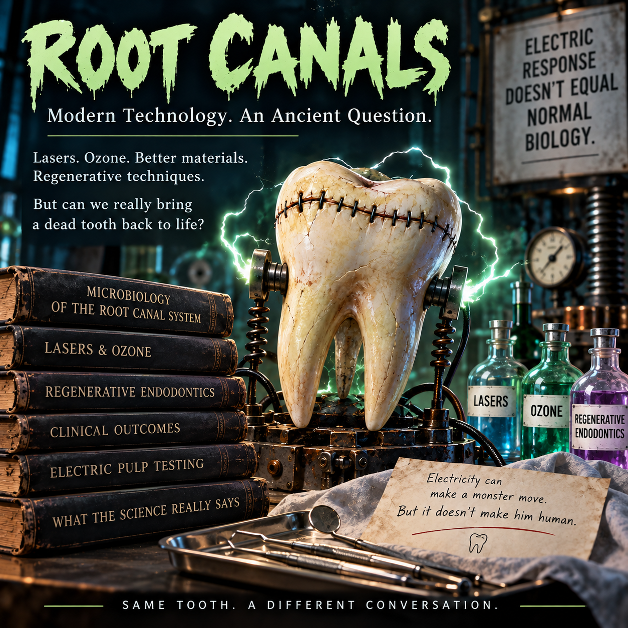
The Root Canal Question: What Are We Actually Saving? Tooth taxidermy, biofilm, zirconia implants, and the human attached to the tooth
Living pulp, tooth taxidermy, protected biofilms, systemic inflammation, zirconia implants—and the human attached to the tooth
A note before we begin: I don't consider conventional root-canal treatment a biologically desirable routine treatment. I recognize that it can reduce infection, relieve pain, and preserve a functional tooth, and that sometimes a root-filled tooth is a necessary temporary compromise—particularly while a young person's jaws develop or an orthodontic or palatal-expansion plan is completed. Those benefits do not make the original internal pulp come back to life or erase the unanswered biological questions. I don't recommend ignoring infection, and I don't believe my personal preference proves that every asymptomatic root-filled tooth should be extracted. This is the distinction that runs through this entire resource.
This is the scientific master behind my ten-part Root Canal Question series. The shorter articles each tackle one subject. Here, I am connecting the entire argument, including the strongest contrary data and the studies I still desperately want somebody to perform. Research, clinical observation, biological plausibility, and hypotheses are not interchangeable; you'll see me label which one I'm discussing.
I went to the IAOMT meeting in Columbus in September 2026, not really excited. It felt like the entire meeting was devoted to root canals—and to be quite frank, a couple of the lectures were PRO root canal. And if you’ve spent approximately twelve minutes around me, you probably already know how I feel about those. So first, I want to say something that sometimes gets lost in conversations about biological dentistry: root canals are still controversial even within the biological dental world. There are really smart, thoughtful people who consider themselves biological dentists and believe root canals can be done safely under the right circumstances.
And then there’s me.
I made a post about it in my biological dentistry group for women dentists. I started a group for female dentists to educate them about biological dentistry about 10 years ago. The discussion that followed from that post was so complex and interesting that I genuinely wish we could offer continuing-education credit for it. I started with a Facebook group post and apparently decided to hold an entire conference in the comments. Needless to say, that conversation turned into another resource-library article for YOU - because that’s how I roll!
Because these are some great questions.
- What does treatment actually preserve?
- What might remain afterward?
- How do we weigh the biology of retaining a tooth against the biology of removing it?
- And what can we honestly tell the human attached to that tooth?
After listening to all of the lectures, I have to tell you I’m still not a fan of root canals. And I don’t believe adding lasers, ozone, different filling materials, or the word “biological” turns a conventional pulpectomy into a treatment that preserves living pulp. When I say there is no such thing as a “biological root canal,” THAT is what I mean: improved disinfection isn't the same as maintaining the original living organ. Better treatment can improve what we accomplish. But it doesn’t automatically resolve the biological objections I’m about to explain. You absolutely deserve more than “because Teresa said so.” And an endodontist (Or ANY dentist, for that matter) reading this deserves better than being told they don’t care about their patients. The conversation that prompted this article happened precisely because we BOTH care. A lot.
So let’s get into it. And yes, I brought the receipts. There are DOZENS of PubMed articles WITH LINKS at the end of this resource-library article. You know me well enough to know I’m not going to write an article built on thin science. We’ve gathered more than 1,600 PubMed links (and GROWING) on this platform because the oral-systemic connection deserves considerably more attention than it gets. AND I’m in the middle of writing a book with more than 2500 references. It’s going to take me the better part of a year to write it because it’s meant to be an amazing resource. There’s a substantial scientific foundation for the questions biological dentistry asks. I intend to SHOW you that science—not just tell you we have it.
And I want the right receipt attached to the right claim. That matters just as much as the size of the reference list. So although this is not intended to be a scientific journal article, I’ve started annotating my articles and references appropriately. My last few articles are written that way. I will eventually rewrite ALL of the articles in this platform in that manner so that you can see exactly how the science relates to the things biological dentistry stands for.
A tooth is a little organ. The pulp isn’t packing material.
A healthy pulp isn’t just “the nerve.” It contains blood vessels, nerves, connective tissue, immune cells, and cells involved in making and repairing dentin. Odontoblasts—the cells along the pulp-dentin border—don’t just build tooth structure. They recognize microbial products and participate in the tooth’s defenses. The pulp responds to injury, regulates inflammation, and participates in repair.[1]
There’s an entire little biological operation happening in there.
Conventional root-canal treatment removes diseased pulp tissue and necrotic contents, cleans and disinfects the canal system as thoroughly as possible, and fills it. The clinician then restores the tooth so the remaining structure can keep functioning. Treatment can relieve pain, maintain mechanical function, reduce the infection but not sterilize the tooth, and allow the tissues around the root to heal.[2,3]
What it CAN’T do is recreate the original living pulp by cleaning the space and putting filling material in it. It doesn’t restore the pulp’s blood supply, original nerve connections, or the living cellular network that coordinates its internal immune responses and repair.[1,55–60].
That’s why I call conventional root-canal treatment “tooth taxidermy”.
A root canal retains the structure and restores useful mechanical function without restoring the original internal living tissue. If the pulp was already necrotic, the disease or injury killed it before treatment. The procedure didn’t cause that original loss. It also doesn’t reverse it. And yes, the periodontal ligament, surrounding bone, and gum tissues remain alive and continue communicating with the body. We’re going to talk about what those tissues preserve because they matter enormously. But the living tissues AROUND the root don’t recreate the missing living system INSIDE it. Keeping the ligament and regenerating the pulp are two different biological accomplishments.[1,34,41]
We’ve preserved some biology. We haven’t preserved ALL the biology. And the missing part wasn’t just sitting there waiting to annoy you.
That’s a much bigger loss than “we took the nerve out so it wouldn’t hurt anymore.” I also consider the tooth-to-organ relationships described in the traditional dental-meridian framework—what I think of as a bioelectrical superhighway. Now, I know the word "meridian" makes some people immediately stop listening, so I want to address that carefully and scientifically later in this article. Keep reading. I promise you will get the science here. Not the woo.
Bioelectrical activity itself isn't controversial. Living cells maintain electrical gradients, nerves transmit electrical signals, and we routinely measure electrical activity in the heart and brain with EKGs and EEGs. The interesting question is what we actually know about how those signals, along with neural, vascular, immune, and connective-tissue pathways, may relate to communication between distant parts of the body. I’ll come back to that in detail later, because the evidence deserves a much more careful discussion than either “meridians are proven” or “meridians are nonsense.”
The nerve isn’t just the tooth’s complaint department.
The pulp’s cells communicate with each other. They respond to microbial signals. Sensory nerves influence immune responses and repair, and immune cells can influence what happens to those nerves. That interaction is called neuroimmune signaling. It isn’t a synonym for pain. It’s a set of biological relationships we can investigate experimentally.[55–60]. And some of this research is REALLY interesting.
The cells that build dentin can also call for immune help.
Durand and colleagues exposed human odontoblast-like cells to lipoteichoic acid, a component of certain bacterial cell walls. The cells responded through microbial-recognition pathways and released chemokines—chemical signals that help recruit immune cells. Fluid collected from the stimulated cells attracted immature dendritic cells, which participate in immune surveillance.[55]
In other words, those tooth-building cells weren’t just producing dentin and minding their own business. They were responding to a microbial challenge and helping organize a defense. Staquet and colleagues then compared odontoblast-like cells with pulp fibroblasts, the connective-tissue cells people might assume are mostly structural. Both responded to microbial signals, but with different patterns of immune messengers. Both could help attract dendritic cells in the laboratory.[56]
That gives us a more interesting picture than “there are some white blood cells in the tooth.” Several kinds of pulpal cells participate in the response.
Odontoblasts can communicate with sensory neurons, too. Shibukawa and colleagues studied odontoblasts alongside trigeminal sensory neurons in experimental rat cell cultures. Mechanical stimulation triggered calcium signaling in the odontoblasts and release of ATP through pannexin-1 channels. That ATP helped transmit signals to neighboring cells and neurons. Blocking particular parts of the pathway interrupted the communication.[57].
You don’t need to memorize the channel names. The important part is that researchers could stimulate a cell, measure a response, and interfere with the pathway to investigate how the message traveled. That’s measured local cell-to-nerve communication. Not a vague claim that teeth “have energy,” and not proof of a tooth-specific pathway to a distant organ.
Nerves and immune cells affect each other—and pain doesn’t tell the whole story.
In a mouse pulp-injury study, Erdogan and colleagues investigated CGRP, short for calcitonin gene-related peptide, a messenger released by sensory nerves. It influenced immune-cell recruitment and pain-related behavior. When the researchers depleted neutrophils and monocytes, they found more sensory-nerve damage and bacteria extending deeper into the tissue—yet lower mechanical pain responses later in the experiment.[58]
That catches my attention. In that model, a smaller pain response didn’t mean better tissue protection. It doesn’t diagnose the person reading this. It shows why I want us to understand the underlying biology instead of treating pain, immune defense, and healing as though they’re the same measurement.
Sensory nerves help direct repair. They don’t just announce the damage.
Moore and colleagues investigated CGRP and another signaling molecule called Sonic hedgehog. Yes, that’s its actual name. Apparently scientists are allowed a sense of humor, too. In mouse injury models and cell experiments, these signals influenced pulp-cell responses associated with expansion and differentiation into dentin-forming cells.[59]
Then Wang and colleagues studied how sensory nerves guide dental pulp stem cells toward an injury. In rat experiments, disrupting the nerve supply impaired stem-cell recruitment and organized repair. CGRP acting through a RAMP1-containing receptor system helped direct those cells, and supplying CGRP improved selected repair responses.[60]
The researchers interrupted a nerve experimentally. That isn’t the same procedure as a clinical root canal. What the experiment helps establish is that sensory innervation participates in coordinating repair, not merely reporting that repair is needed. Now put those findings together. The pulp contains cells that detect microbial products, release immune messengers, communicate with sensory neurons, and respond to signals that help organize tissue repair. Different experiments establish different parts of that picture.[55–60]
Calling the pulp “the nerve” leaves out an awful lot of what we’re trying to preserve. And once you understand how much communication is actually happening inside a living pulp, the next question becomes pretty obvious: what exactly replaces all of that when we remove it?
A filling doesn’t replace a cellular conversation.
That’s the biological point I don’t want buried beneath a discussion about how beautifully the canals were cleaned. A conventional root filling doesn’t become a population of odontoblasts, fibroblasts, immune cells, and sensory endings working together inside a vascularized pulp. A material may interact with tissues, and a treated tooth may function well, without recreating that system.[1,2,55–60]
We also shouldn’t confuse the absence of the original pulpal immune network with the absence of ALL immune activity around the tooth. The surrounding tissues can still respond. That’s part of how periapical disease develops and how it heals.[1,5]
For me, this is a powerful argument for preserving healthy or recoverable pulp whenever reasonably possible. It also raises the standard for what we should eventually call successful pulp regeneration: not just tissue occupying a space, but appropriate tissue doing the jobs it belongs there to do. And regardless, you need to understand this: once the pulp is completely necrotic, those normal functions are already lost. Neither filling the tooth nor extracting it brings them back. Choosing between those options still requires the infection, structural prognosis, supporting tissues, replacement plan, and human being to be assessed together.[1,2,5]
So this research doesn’t make extraction the automatic answer. It makes “the tooth is still there” an incomplete description of what treatment accomplished. That’s not a small distinction to me.
Before we remove the pulp, can we save the part that's STILL ALIVE?
When people hear me criticize root canals, a perfectly reasonable question follows: "Okay, Teresa, but what if the nerve is hurting and I don't want to lose my tooth?" The first answer should not be to assume the pulp must come out. The first answer should be to determine whether there is living tissue we can preserve. And I mean LIVING tissue—not a tooth that happens to be sitting there after its original pulp has died.
Vital pulp therapy, or VPT, only preserves living pulp that is still present. If the ENTIRE pulp has already become necrotic, or has been removed in a conventional root canal, ordinary VPT cannot bring that original organ back. I need that sentence to be unmissable. I do not want somebody with a completely necrotic, infected tooth spending months searching for a pulpotomy that cannot achieve what they're imagining. Please don't confuse genuine tissue preservation with tissue regeneration, which is a very different and still developing field.[2,92,93]
There is an important anatomical nuance, though. A tooth can have severely inflamed or necrotic coronal tissue while deeper radicular pulp remains alive. If an adequately healthy, vascularized portion of pulp is still present, a carefully selected pulpotomy may preserve it. "Some pulp is necrotic" is not necessarily equivalent to "the entire pulp is necrotic." On the other hand, cold-test silence alone does not give me permission to label the entire pulp dead, and pain alone does not prove it's all hopeless. Proper diagnosis, clinical examination, imaging when indicated, and sometimes direct assessment of tissue and bleeding are essential.[2,39,41,92,93]
Four procedures that do NOT remove the same amount of pulp
Indirect pulp treatment manages deep caries while avoiding intentional pulp exposure. Direct pulp capping covers a limited exposure in a tooth judged capable of healing. Partial pulpotomy removes a limited portion of damaged coronal pulp, aiming to retain healthier coronal and radicular tissue. Full coronal pulpotomy removes the coronal pulp but preserves the living tissue inside the root canals. That last approach is particularly easy to misunderstand. The chamber is NOT left empty after the procedure: a suitable material protects the remaining pulp at the canal entrances, and an adequately sealed restorative build-up and definitive restoration close the space above it. The radicular pulp remains living tissue, rather than being removed and replaced by a conventional root filling.[2,92,93]
The science behind selecting the viable portion is more than wishful thinking. Ricucci and colleagues studied 264 human teeth histologically and histobacteriologically, along with a separate clinical series of 757 VPT cases. They demonstrated that inflammation and bacterial invasion in carious teeth may be localized rather than uniformly distributed throughout every millimeter of pulp. In their clinical series the reported success rates differed substantially by procedure. The exact technique, diagnosis, patient selection, restoration, and long-term observation matter; I wouldn't apply one percentage indiscriminately to every tooth.[92]
And I want to be clear that less tissue removed is not automatically safer. If inflammation has extended beyond the portion being removed, a partial pulpotomy may preserve more tissue in theory but leave an unacceptably compromised environment in practice. A 2026 synthesis of 23 studies of mature permanent posterior teeth with irreversible pulpitis found pooled 12-month success of about 92% for full pulpotomy and 89% for partial pulpotomy. Direct comparisons favored full pulpotomy for short-term success and pain-related failures, although the certainty of evidence was low to very low and the longer-term comparisons were much thinner. I value preserving biology. I also want the tissue that remains to be genuinely capable of surviving.[103]
What does successful vital pulp therapy ACTUALLY mean?
I want to establish something right here because I think the way dental research reports success can be terribly misleading. When I evaluate vital pulp therapy, I have ONE primary biological objective: I want the original pulp tissue we've preserved to remain ALIVE.
Not just for six months. Not just for a year. I want to know whether that pulp is still alive at one year, three years, five years, ten years, and twenty years. That's what we're trying to accomplish!
If we perform a pulpotomy and the remaining pulp subsequently becomes completely necrotic, the vital pulp therapy has failed to preserve the living biology we were trying to save. If that tooth then requires a conventional root canal, the VPT has failed its biological objective. The patient may still have a functional tooth, but we've lost the original living pulp. Its vascular supply, internal immune network, and original pulpal neuroimmune communication are no longer there.
And I REFUSE to call that successful preservation of vitality simply because the tooth hasn't been extracted. There's a difference between tooth survival, clinical success, and continued pulp vitality. Those are separate outcomes, and I want the research to report them separately.
Interestingly, Chien and colleagues published a 2026 review specifically examining what we mean by success in vital pulp therapy. They found substantial inconsistency in how outcomes are defined and measured across studies. They also explained that commonly used indicators, including sensibility testing, symptoms, and radiographic findings, don't necessarily give us a complete picture of the pulp's biological condition.[105]
Now THAT deserves attention.
Because if the entire purpose of VPT is preserving living pulp, shouldn't continued pulp vitality be the primary biological outcome we're measuring? I certainly think so.
Of course, demonstrating that isn't always straightforward. Cold and electric pulp testing primarily measure sensory responsiveness rather than directly measuring blood circulation. Laser Doppler flowmetry and pulse oximetry can investigate vascular vitality more directly, although both have limitations in routine clinical application.[41,100]
But the limitations of our testing shouldn't change the biological objective we're trying to achieve. I want the researchers to distinguish between teeth with evidence supporting continued vitality, teeth that have definitively lost their pulp, and teeth whose vital status remains uncertain. And I want those outcomes followed for DECADES. Not the nonsense 6 and 12 month studies.
How long can vital pulp therapy actually keep the pulp ALIVE?
This is the question I really want answered. Because the entire purpose of vital pulp therapy is to preserve the LIVING tissue inside the tooth.
I don't want to know simply whether a tooth remains functional. I want to know whether the original pulp tissue we intended to preserve is STILL ALIVE at one year, three years, five years, ten years, and twenty years. That's my definition of successful vital pulp therapy.
A 2025 systematic review reported approximately 91–97% clinical success at twenty-four months across several VPT procedures in appropriately selected permanent teeth initially diagnosed with normal pulp or reversible pulpitis.[94]
That sounds encouraging, but there's an important distinction. Those percentages reflect the clinical success criteria used in the included studies. They don't necessarily establish that the remaining pulp was directly confirmed to have a functioning blood supply, appropriate immune activity, and preserved neurological function.
And that's the biological outcome I'm interested in.
Eggmann and colleagues evaluated 64 eligible partial-pulpotomy cases over an average follow-up of approximately 3.1 years. Fifty-seven met the study's criteria for successful pulp preservation, giving them a reported success rate of 89.1%.[95]. But let's look at what happened to the other seven teeth.
Six subsequently required conventional root-canal treatment. That means the original pulp tissue we were trying to preserve was ultimately removed. Those six cases therefore failed to achieve the biological objective of VPT. The seventh tooth fractured and was extracted. That's a different complication, and the fracture alone doesn't establish whether the pulp was still alive before extraction.
Here's what matters to me: Six patients underwent a procedure specifically intended to preserve their living pulp, but ultimately lost that pulp and required conventional root-canal treatment. I don't consider those six cases successful vital pulp therapy.
Now, that doesn't mean the treatment accomplished nothing while the pulp remained alive. If we preserved living tissue for three years before it eventually failed, those years of preservation had value. But three years of maintained vitality isn't the same thing as ten years of maintained vitality. And once that original pulp is completely necrotic or has been removed, the biological objective has been lost.
A five-year follow-up of a randomized clinical trial examined partial and full pulpotomies in mature permanent molars diagnosed with symptomatic irreversible pulpitis. Among the 59 participants evaluated at five years, approximately 83% met the study's combined clinical and radiographic criteria for success.[96]. Again, that's encouraging. But I want to know how many of those teeth still contained living, vascularized pulp tissue.
The investigators found that partial pulpotomy produced more positive cold-test responses and less pulp canal obliteration than full pulpotomy. Those findings are interesting because they may reflect differences in preserved sensory function and tissue responses. But a cold-test response isn't a direct measurement of pulpal blood circulation. And an absence of symptoms or radiographic disease doesn't establish that the pulp remains alive. So while these studies provide evidence that VPT can achieve favorable clinical outcomes for years, they don't always answer my most important question with the biological precision I'd like.
How much of the original living pulp remains alive, and for how long?
That's what I want researchers to investigate. I want studies that directly assess continued pulp vitality wherever feasible, distinguish confirmed vitality from uncertain vitality, and follow those outcomes for DECADES.
Because successful vital pulp therapy, as I'm defining its primary biological endpoint, means that the living pulp we intended to preserve is STILL LIVING. Period.
Now, let's look a little further down the road. A five-year follow-up of a randomized clinical trial examined partial and full pulpotomies in mature permanent molars diagnosed with symptomatic irreversible pulpitis. Of the 59 participants evaluated at five years, approximately 83% met the study's combined clinical and radiographic criteria for success.[96]
That's encouraging, particularly because these weren't simply young children's teeth with incomplete root development. They were mature permanent molars with significant pulpal disease.
But there are two details I want you to notice.
First, only 59 of the original 93 participants were evaluated at five years. We can't automatically assume that everyone who wasn't evaluated had a successful or unsuccessful outcome.
Second, partial pulpotomy produced more positive cold-test responses and less pulp canal obliteration than full pulpotomy. That's interesting because preserving more of the original tissue may also preserve more of its sensory responsiveness.
But an electric or cold response isn't a direct measurement of pulpal blood flow. And pulp canal obliteration doesn't automatically establish that the remaining tissue has died. So while these findings provide encouraging evidence of long-term clinical success, they don't prove that every tooth retained completely normal pulp biology throughout those five years.
And that's what I want future research to investigate.
Kunert and colleagues also retrospectively analyzed 273 pulpotomies, with observation periods ranging from one to twenty-nine years.[98]. That tells us some treated teeth can remain clinically successful for a very long time. It does NOT mean every tooth was followed for twenty-nine years, nor does it mean researchers directly verified continued pulp vascularity throughout that entire period.
I want to know whether those teeth still contained living pulp at five years, ten years, fifteen years, and twenty years.
And I want that answer separated from whether the tooth had subsequently received a conventional root canal or simply remained present in the patient's mouth. Because if the tooth required complete pulpectomy somewhere along the way, we didn't successfully preserve its original living pulp for that entire period.
That doesn't erase the years of vitality the patient may have benefited from before the failure. Those years had value. But we need to report how long vitality was actually maintained rather than counting the subsequent root-filled tooth as continued VPT success.
And in growing children, those years can be especially valuable.
A 2026 retrospective study examined 192 immature permanent teeth in 108 young patients who underwent cervical pulpotomy. The investigators reported 91.7% clinical success over an average follow-up of approximately 63 months.[97]. These were selected teeth with living pulp at the time of treatment, not teeth whose entire pulp had already become necrotic. And the biological advantage in an immature permanent tooth can be particularly important.
When living radicular pulp and the associated developing tissues remain functional, continued root maturation may occur. That's called apexogenesis. We're not merely trying to keep a tooth-shaped object in a child's mouth until the jaws finish growing. We're trying to preserve the living biological processes that allow that tooth to continue developing. And when a child needs orthodontic treatment, certain types of palatal expansion, or other growth-related treatment, preserving a strategically important living tooth may provide additional benefits.
But I still want the same biological question answered: Is the pulp STILL ALIVE? If the pulp remains alive and healthy, we've preserved something meaningful. If it becomes completely necrotic and the tooth requires a conventional root canal, we've lost the defining biological benefit of VPT, even if retaining the tooth remains strategically necessary for the child's treatment. Those are two different accomplishments, and they shouldn't be confused.
Here's the research I actually want.
- I want a long-term prospective study that follows patients after vital pulp therapy at one, three, five, ten, fifteen, and twenty years.
- I want researchers to document the original diagnosis, how much pulp was removed, which materials were used, and what evidence demonstrated that the remaining tissue was vital.
- Then I want them to measure continued pulp vitality at every follow-up using the best clinically appropriate methods available.
- I want tooth survival reported separately.
- I want the need for subsequent conventional root-canal treatment reported separately.
- I want recurrent decay, restorations, fractures, radiographic changes, pulp canal obliteration, and other complications documented.
- And when pulp vitality cannot be determined confidently, I want the researchers to acknowledge that uncertainty rather than automatically counting an asymptomatic tooth as biologically successful.
A tooth that still responds to cold isn't automatically proven to have completely normal pulp biology. But a tooth that has undergone complete pulpectomy definitively no longer contains the original living pulp we intended to preserve.
And that is a distinction we CAN make.
My definition of successful vital pulp therapy is straightforward: The original pulp tissue we intended to preserve remains alive.
- At one year.
- At three years.
- At five years.
- At ten years.
- At twenty years.
- And ideally, for the rest of that tooth's useful life.
That's the biological outcome I want us working toward. Because my objective isn't simply to keep a tooth in someone's mouth. My objective is to keep the living biology INSIDE that tooth alive whenever we reasonably can. THAT is what successful vital pulp therapy means to me.
And YES, the material over the pulp matters
Calcium-silicate cements are commonly used because of their favorable clinical and tissue-response profile, but materials and methods are not interchangeable. In a 2024 network meta-analysis of 21 trials, calcium hydroxide showed greater failure odds than MTA at several time points; selected other calcium-silicate materials did not show statistically significant differences from MTA in the available comparisons. That does not mean every branded product has identical resin additives, radiopacifiers, setting chemistry, exposure profiles, or patient compatibility.[99] Some published long-term series used older material systems. We should not hide that just because their survival data look encouraging.
The 2025 permanent-tooth VPT guideline addresses selection, bleeding control, calcium-silicate materials, and the need to assess pulpal status directly in difficult cases. It generally recommends full rather than partial pulpotomy for certain deep-caries presentations with spontaneous, nocturnal, or lingering pain once the pulp has been determined to be vital and controllable. It does NOT turn a tooth with a completely necrotic pulp into a VPT candidate.[93]
Buying time for growth is different from restoring vitality
Some children or adolescents need strategically important teeth to maintain function, space, or anchorage while their jaws mature or orthodontic and palatal-expansion plans proceed. Certain tooth-supported expansion methods depend on teeth; other expansion methods do not. And the ability to achieve meaningful airway change is not determined by tooth retention alone. I don't want people turning a real clinical consideration into a universal statement that "you can't grow airways without teeth." The correct question is what THIS child needs for THIS treatment plan.[51,52]
If a tooth still has healthy, preservable pulp, VPT may protect living tissue AND maintain the tooth during growth. If the pulp is fully necrotic, VPT is not an option. Conventional root-canal treatment might sometimes become a carefully monitored temporary strategic compromise when extracting a key tooth would seriously undermine growth-related or orthodontic treatment. But that does not make conventional root filling the biological goal, and it does not justify ignoring an uncontrolled infection. When the strategic reason for retention has passed, the patient deserves an updated discussion of risk, prognosis, and options.[2,51,52]
A failed pulpotomy also needs reassessment. Remaining pulp may become necrotic; secondary caries, leakage, missed pathology, and structural problems can change the prognosis. When that happens, the ORIGINAL biological advantage has been lost. We then need a fresh diagnosis rather than continuing to call a tooth vital just because somebody performed VPT on it years earlier.
My position is straightforward: if we can truly preserve living pulp, investigate that first. If the entire pulp is already dead, do not promise VPT. And if we're temporarily retaining a nonvital tooth for an important developmental reason, call it what it is—a compromise with a purpose, not a restoration of lost biology.
Before anybody decides to keep squeezing an abscess… NO.
Root canal INFECTION and root canal TREATMENT aren’t the same thing. I don’t ever want someone reading this and thinking, “Well, Teresa doesn’t like root canals, so I’ll just leave this dead, infected tooth sitting here and periodically squeeze the pimple on my gum.” NOOOOOOO!!! That pimple on your gum may be a draining sinus tract. Drainage can relieve pressure without eliminating the source of infection. A necrotic infected tooth needs assessment and treatment.[2]
My disagreement isn’t about whether we should treat infection. It’s about what treatment leaves behind, what it sacrifices, and which option makes the most sense for the person receiving it. A root filling, by itself, isn’t a diagnosis of active infection. An untreated abscess, a root-filled tooth with persistent apical disease, and a root-filled tooth with healed surrounding tissues aren’t interchangeable situations.[2,5]. At the same time, when infection persists or comes back, I don’t consider it less important because somebody already performed a root canal. The tooth’s treatment history doesn’t exempt its current biology from investigation.
The problem isn’t that endodontists don’t know how to clean a tube. It’s that a tooth isn’t a tube.
When people picture a root canal, they usually imagine a hollow drinking straw running down the middle of a root. If that were the anatomy, this would be a much shorter article. Instead, we have main canals, lateral canals, accessory branches, fins, isthmuses connecting spaces, apical ramifications near the root tip, and microscopic dentinal tubules running through the dentin. Instruments can shape the larger spaces. They can’t physically travel through every branch and tubule.[4,5]
Irrigants can reach beyond where instruments touch. Activating those irrigants can improve their movement and contact with surfaces. But reaching farther isn’t the same as reliably reaching EVERYTHING. The endodontic literature has been describing this problem for decades. Wu and colleagues explained why conventional sampling can miss organisms outside the main canal. Histologic research has shown organized biofilms in diseased root-canal systems. Other investigators have combined microbiology, three-dimensional imaging, and histology to examine where infection persists after treatment.[4–7].
This isn’t biological dentistry making up anatomy because we don’t like root canals. This is endodontic research.
And it matters even when the clinician is excellent. Some problems are caused by poor treatment. Others are caused by anatomy that remains difficult to disinfect despite excellent treatment. Those aren’t the same accusation. A biofilm also isn’t just a few lonely bacteria floating around waiting for disinfectant to arrive. It’s an organized microbial community living in a matrix of material the organisms produce, interacting with its neighbors, and behaving differently from free-floating organisms. The same chemical can perform differently against those two situations.[6,19]
So my concern isn’t merely, “Did we kill some bacteria?” Of course we did. My concern is: what kind of habitat remains for whatever survived, and what keeps that habitat from becoming a problem later?
What I’ve actually found in the teeth I’ve tested
Every single root-canaled tooth I’ve personally extracted and sent for microbial DNA testing has come back with a microbial report that made me uncomfortable.
Every. Single. One.
Usually, we aren’t talking about one organism. We’re talking about a list. And I’ve seen organisms—including Legionella species—that I don’t routinely see in the periodontal samples I submit. That’s MY clinical experience. I’m not going to erase it because it didn’t arrive wearing a randomized-trial name tag. I’m also not going to claim those reports answer questions they don’t answer. These are teeth that were extracted and tested, not a random sampling of everyone walking around with a root canal. DNA testing doesn’t prove every detected organism was alive when the tooth came out. A long list isn’t automatically a measurement of live bacterial quantity. And an unusual finding deserves confirmation rather than an automatic conclusion about where it came from.
When I call this pattern a “biofilm bloom,” I’m describing my clinical interpretation and my concern about microbial persistence or re-establishment. The panel doesn’t photograph a biofilm or tell me when it grew. I can be clear about that without pretending the pattern means nothing to me. Published research gives that concern a biological basis. A 2023 scoping review of 32 clinical studies described persistent endodontic infections as polymicrobial. Organisms included Enterococcus faecalis, Parvimonas micra, Porphyromonas endodontalis, Prevotella intermedia, and others. Histologic work documents protected microbial communities, not simply an empty canal that happens to have an unfortunate-looking laboratory report.[6–8]
That doesn’t make Legionella an established signature of root-canal infection. My observation and the published evidence are different pieces of information. I want readers to know which one I’m discussing. My experience has formed my clinical judgment. I won’t tell every person with a root canal that they’re being poisoned. But I’m also not going to tell every person with a root canal that they are fine. We aren’t cookie cutters. Every person’s immune system is different. I expect to treat every person with their own body’s considerations. I would hope that all doctors and dentists do the same for their own patients. Alas, conventional medicine tries to fit people into boxes. This is a habit I’m trying to break.
Here’s a study that deserves more attention
Hernández and colleagues examined 45 extracted root-filled teeth. Twenty-three had apical periodontitis—inflammation around the root tip—on cone-beam CT imaging, or CBCT, and twenty-two didn’t. They examined the root tips using complementary laboratory methods and detected bacterial DNA in 44 of the 45 specimens. Mean total bacterial counts didn’t differ significantly between the groups.[9]
Read that again: the group WITHOUT a visible apical lesion wasn’t automatically the group without bacterial DNA. Now, the authors described relatively low mean counts. These weren’t 45 randomly selected people, and this wasn’t proof of systemic illness. But it’s an important reason not to confuse two different questions: “Does the scan show an apical lesion?” and “Is microbial material detectable inside the apical canal?”
A scan isn’t a microbial census. And a microbial census isn’t a whole-person diagnosis. For me, that’s exactly why the next question matters: what determines whether a particular microbial situation is clinically important for a particular human?
“But the bacteria might be dead.” Yes. Let’s actually examine that.
Ordinary DNA testing can detect genetic material from organisms that are no longer alive. That’s a legitimate limitation, and I’m not arguing with it. But when someone tells me, “The bacteria might be dead,” my next question is: Okay. WHEN did they die?
Before treatment? During disinfection? While the tooth was still in the patient? After extraction? During collection? During transport? While somebody was processing the specimen in the laboratory? A DNA-only report doesn’t answer that. And “we haven’t proven they were alive” is NOT the same statement as “we’ve proven they were dead before the tooth came out.”
Research actually demonstrates why that distinction matters. Brundin and colleagues deliberately exposed the anaerobic organisms Fusobacterium nucleatum and Peptostreptococcus anaerobius to air until they lost viability. Their DNA remained detectable by conventional PCR for SIX MONTHS afterward.[64]. That matters to me because anaerobic bacteria are, by definition, organisms whose survival may be affected by oxygen exposure. So if an organism is alive inside a low-oxygen environment and loses viability after it’s removed, the later DNA result can’t reach backward in time and tell me exactly when that death occurred. But—and this is important—the uncertainty runs BOTH ways.
A separate Brundin study put already heat-killed Enterococcus faecalis into extracted human root canals. Amplifiable bacterial DNA could still be detected one and two YEARS later.[65]. So yes, a positive DNA test may represent an organism that died during treatment or long before the tooth was removed. It may also represent an organism that was viable closer to the time of extraction and died afterward.
The DNA test doesn’t come with a microbial death certificate. Or a timestamp.
Even conventional culture is affected by what happens AFTER the specimen is collected. Carlsson and Sundqvist compared different transport and cultivation methods for root-canal specimens and recovered bacteria from anywhere from 29% to 58% of samples depending on the method used.[66]. That’s a pretty enormous swing. It doesn’t mean culture is useless. It means a negative culture can’t automatically prove an organism wasn’t viable before collection when the collection, transport, oxygen exposure, media, and laboratory conditions themselves affect what ultimately grows.
And I want to be equally careful in the other direction. Finding microbial DNA inside an extracted tooth does NOT prove that every organism on that report entered that patient’s bloodstream or caused systemic disease. The tooth specimen doesn’t establish that.
But bloodstream entry from endodontic procedures isn’t imaginary, either. Savarrio and colleagues actually looked for it during nonsurgical root-canal treatment. They detected bacteremia by conventional culture in 9 of 30 patients. In seven patients, the same bacterial species was identified in both the root canal and bloodstream, and in the two pairs they genetically typed, the isolates were identical.[67]
That demonstrates that endodontic organisms CAN reach the bloodstream.
It does NOT give me permission to say that every organism on every extracted-tooth DNA report circulated through that patient’s body. Possibility and proof in a particular person are two different things. That’s exactly the kind of distinction I want biological dentistry to be GOOD at making. So yes, when somebody says, “But those bacteria might be dead,” my answer is: Absolutely. They might be. But this test can’t tell me WHEN they died. And nobody gets to use that uncertainty in only one direction. I can’t honestly claim every organism on a DNA report was alive inside the patient at the moment of extraction. But nobody else can honestly use that exact same DNA-only report to claim those organisms were already dead and biologically irrelevant while the tooth was still in the patient.
The limitation of the test is a reason to be precise about the result. It isn’t proof of danger, and it isn’t a certificate of harmlessness.
And there’s another detail that gets muddled even in professional conversations because, in all fairness, most of us—including me—aren’t molecular microbiologists: 16S rRNA GENE sequencing usually examines DNA. The “RNA” in the name doesn’t magically turn it into a test of living bacteria. Testing actual RNA requires a different laboratory process.[9,11,13]
Researchers have better ways to investigate activity. In one clinical study, propidium monoazide combined with quantitative PCR—a method that amplifies and measures targeted DNA—helped distinguish signals associated with intact cells from those associated with damaged cells. It improves the picture, although membrane integrity still isn’t a perfect definition of viability.[10]
Metatranscriptomics goes after RNA to investigate what genes organisms are expressing. A 2024 endodontic study found expressed genes associated with potential virulence and antibiotic resistance. It was small—eight samples yielded enough RNA for full analysis—but it shows why asking what microbes are DOING can be more informative than asking only which names appear on a report.[11]
RNA still needs interpretation. Organisms differ, dormancy complicates things, and detecting ribosomal RNA isn’t a universal “definitely alive and causing disease” stamp.[13]. I’m interested in better testing. Absolutely.
But none of these tools gives me a routine chairside way to sample every dentinal tubule, branch, isthmus, and possible external-root biofilm in an already filled tooth. We can sample during treatment, retreatment, or surgery. We can examine extracted specimens more extensively. Every approach has access limitations.[4,7,9]. And by the time I can send the entire tooth to the laboratory, I’ve already taken it out. There’s the conundrum. And then there’s the cost. My patients currently spend about $450 per sample for the DNA ConneXions report. Insurance certainly doesn’t cover that.
Also, bacterial products don’t disappear just because the conversation switches to viability.
Gomes and colleagues studied 50 teeth requiring retreatment that had radiographic apical periodontitis. They used culture, molecular methods, and separate tests for lipopolysaccharides and lipoteichoic acids—bacterial components relevant to inflammation. Bacteria were detected in all samples by culture and molecular methods. Both types of bacterial products were also detected in every sample. Higher lipoteichoic-acid levels were associated with larger radiolucent areas.[12]
Those were diseased teeth, not a demonstration that every successfully treated tooth contains a harmful dose. But they make it impossible to reduce the entire biological question to whether a particular DNA signal came from a living organism. Viable organisms, microbial products, biofilm architecture, and the human response are related questions. They aren’t interchangeable tests.
My DNA connexions panel doesn’t measure all of them. Saying “DNA doesn’t prove viability” doesn’t make all the other questions go away, either.
We’re ecosystems. That’s a reason to ask MORE questions, not fewer.
I agree wholeheartedly that looking for one bad bug and trying to kill it isn’t always enough. We need to ask why the environment allowed disease to develop, how organisms interact, and how the patient’s biology participates. There’s fascinating science behind this. In an experimental periodontal model, very small numbers of Porphyromonas gingivalis could change the resident microbial community and promote inflammatory bone loss through interactions involving the host’s complement system, part of its immune defenses.[14]
That was a mouse periodontal experiment, not a demonstration of what happens in every root canal. Its lesson is still important: a small count doesn’t necessarily mean a small biological effect. Community behavior and host interaction matter. And those familiar periodontal organisms aren’t harmless just because they already live somewhere else in the mouth. Location, activity, community, and the condition of the surrounding tissues matter. That’s why I don’t divide microbes into “scary root-canal bugs” and “perfectly friendly implant bugs.”[8,14,27,28]
The ecology matters. The species matter. The inflammatory products matter. The host matters. Which brings me to the problem I still can’t get around.
The 86%-17% problem
The strongest argument for keeping a treated tooth sounds something like this: clean it as thoroughly as possible, seal it well, preserve the periodontal ligament, support the patient’s health, and monitor the surrounding tissues. Perhaps we don’t need complete sterility if treatment changes the environment enough for healing to occur.[2–5]
I understand that argument. It’s not a stupid argument. It’s the reason a thoughtful endodontist can disagree with me without either of us needing to declare the other person irresponsible. But when “strengthen the host” becomes the answer to “How much burden can THIS human tolerate from THIS tooth?” I still have a problem. Humans don’t come with little biological-reserve gauges on their foreheads.
“Excellent! You’re currently operating at 86% biological reserve, and I’ve calculated that this root canal will require approximately 17%. You’ve got plenty to spare. Proceed!” I WISH I could do that.
Of course medical history matters. Blood-sugar control matters. The periodontal condition, the tooth’s structure, the quality of treatment and restoration, and the follow-up findings all give us useful information. Research shows that diabetes is associated with worse periapical findings in root-filled teeth.[2,3,15]
We aren’t practicing blindfolded. But none of that gives us a complete measurement of this person’s reserve, everything remaining in that tooth, or the way those things will interact five or fifteen years from now. Optimizing the patient’s health is part of the answer. It isn’t a measurement of the answer. And no, uncertainty doesn’t automatically make extraction safer—or justify extracting a comfortable, functional tooth with no established disease. I have to compare actual alternatives, including their known complications. My point is that “the patient looks healthy” isn’t enough to settle the biological question for me.
He was handling it. Until he wasn’t.
One case in our discussion on the aforementioned Facebook-group discussion involved a tooth whose apical lesion initially improved and later enlarged. In the meantime, the patient had lost his job, spent months looking for work, and was dealing with his marriage falling apart. The proposed explanation was that his immune response had changed and he could no longer maintain the earlier balance. That’s plausible. It isn’t something the case proves. Persistent infection, missed anatomy, leakage, fracture, extraradicular infection, and other causes still belong in the investigation. Initial bone fill doesn’t establish that everything inside the root was adequately eliminated forever.[5]
But the case perfectly illustrates why I keep asking about the human’s future, not just the tooth’s current photograph. He appeared to be doing well. Then the picture changed. That doesn’t mean his earlier treatment accomplished nothing. It means early improvement and permanent biological stability aren’t the same promise. This applies to EVERYBODY. It isn’t that biological dentists have some special collection of medically complicated humans while conventional dentists treat a different species. Hidden and changing health problems don’t check the office sign before entering.
I’m a stage IV cancer survivor. I know what it feels like to have life divided into before and after. That experience affects how seriously I take uncertainty. It isn’t evidence that a root canal caused my cancer - because I have NEVER had a root canal, and I’m not presenting it that way. It’s why this question matters so much to me: What happens if tomorrow’s human has to carry yesterday’s dental decision?
Your immune system needs regulation. It doesn’t need a permanent bar fight.
Yes, humans evolved in relationship with microbes. Yes, a healthy microbiome matters. No, that doesn’t establish that keeping a chronic endodontic infection is useful immune exercise. The immune response itself can contribute to tissue destruction when inflammation doesn’t resolve. The goal isn’t simply “more immunity.” It’s effective defense, appropriate regulation, and healing.[1,5,14]
My immune system probably does need CrossFit. My personal idea of exercise leans heavily toward surfing on couches. But I’m not convinced it needs to wrestle a crocodile in my mandible for twenty years. I want to support the human AND address the source of disease. Those aren’t competing philosophies. “Improve the host” shouldn’t become an excuse to stop examining the habitat the host is being asked to manage.
The inflammatory-marker studies actually strengthen the oral-systemic conversation.
Here’s a point I’m happy to give endodontics: treating apical periodontitis can be followed by reductions in circulating inflammatory markers. A 2025 systematic review included 16 studies and 596 patients; six studies contributed to the meta-analysis. The pooled findings showed a significant decrease in high-sensitivity C-reactive protein, or hs-CRP, at six months—from approximately 2.26 to 1.28 mg/L—and a significant decrease in IL-1β at twelve months.[16]. That’s GOOD.
I WANT the inflammatory burden to go down when we treat an infection. But here’s where my brain immediately goes: “Okay, so what happens NEXT?” What happens AFTER twelve months? Because twelve months is not long-term follow-up for a treatment we’re asking somebody to carry around for ten, fifteen, or twenty years. What do those same inflammatory markers look like at 24 months? Thirty-six months? Five years? Eight years? Ten years? Six and twelve months tell me that we successfully reduced the inflammatory burden associated with the original apical disease during the period that was actually studied. They do NOT tell me that the reduction stays there for the next decade. And that matters when we look at how long root-canaled teeth actually remain in the mouth. Because remember the story mentioned just above - he was fine, until he wasn't.
One enormous real-world study looked at 71,283 root-canal-treated permanent teeth in 46,702 patients across 99 general dental practices. The overall median survival time was 11.1 years, and only about 26% remained event-free beyond twenty years.[68]. At the 11.1-year median, approximately half had reached the study’s defined survival endpoint. Now, before somebody comes for me, that does NOT mean half of those teeth had recurrent apical infections by 11.1 years, and it does NOT mean half had simply fallen out or been extracted. Their survival endpoint included subsequent events such as retreatment, apical surgery, root amputation, extraction, and replacement. The restoration mattered tremendously too. Teeth that received both a filling and a crown had substantially longer median survival.[68]. But that actually makes me MORE interested in the biology, not less.
Because if we’re going to celebrate a measurable systemic inflammatory improvement at six or twelve months while asking that tooth to remain in somebody’s body for another decade or two, I want to know what happens between those two timelines. I want the three-year bloodwork. The five-year bloodwork. The ten-year bloodwork. And I want those blood samples paired with CBCT findings, symptoms, clinical examination, microbial data, changes in the patient’s medical health, AND what eventually happened to the tooth.
And here’s the really interesting part: The inflammatory markers don’t necessarily behave like a nice little line that simply goes down after treatment and stays there.
Bakhsh and colleagues followed patients undergoing nonsurgical root-canal retreatment or periapical surgery for apical periodontitis. They measured a collection of circulating inflammatory biomarkers before treatment and again at three, six, and twelve months.[69]. And the pattern was COMPLICATED. At three months, IL-1β, IL-8, hs-CRP, complement C3, MMP-2, and MMP-9 were higher than baseline, although not every increase was statistically significant in every treatment group. At six months, IL-1β and IL-8 increased further while several other markers decreased. By twelve months, some markers had fallen below baseline, others were closer to baseline, and the pattern still wasn’t one simple downward trajectory.[69]. Not every marker had fallen below baseline at twelve months. The authors interpreted some of the earlier increases as responses to treatment and remodeling, which is plausible. But selecting one favorable biomarker while ignoring a mixed panel would be cherry-picking in either direction. I want the whole panel, not just the portion that supports a preferred story.
But that actually proves MY point: the biology is considerably more complicated than “root canal done → inflammation goes down.”
This part really bears paying attention to: At the one-year CBCT evaluation, only 25.9% of the treated cases were classified as completely healed. Sixty-three percent were still healing, and 11.1% had failed. Six-month IL-8 levels were associated with the eventual twelve-month treatment outcome.[69]. In other words, an inflammatory biomarker measured BEFORE the final CBCT outcome contained information related to how that case eventually turned out. Does that prove inflammatory markers start changing years before every root canal fails on an X-ray? No.
But holy cow, doesn’t it make you want to LOOK?
Because I do. And we already know that apical periodontitis itself is associated with systemic inflammatory changes. A 2026 study of 206 adults found that more severe apical periodontitis was associated with higher systemic immune-inflammatory index, neutrophil-to-lymphocyte ratio, and platelet inflammatory value.[72]. So there’s a measurable relationship between the severity of what’s happening around the root and inflammatory status in the human attached to it. Then another 2026 systematic review looked specifically at whether endodontic treatment changes serum inflammatory markers in people with asymptomatic apical periodontitis. It found possible reductions in CRP, IL-6, and TNF-α after treatment—but the certainty of that evidence was rated very low.[71]
And a prospective 2024 study of young, clinically healthy adults with apical periodontitis found reductions in hs-CRP and monomeric CRP at one and six months in the subgroup that began with elevated cardiovascular-risk hs-CRP. But TNF-α, IL-6, IL-10, IL-1β, and soluble E-selectin didn’t significantly change.[70]. Again: biology is not giving us one tidy number. So when somebody says, “See? Inflammatory markers went down after the root canal,” my answer is:
Great. I’m genuinely glad they did. Now show me YEARS THREE, FIVE, EIGHT, and TEN. And show me ALL of the inflammatory markers—not just the ones that went down.
Because THAT’S the study I want.
- I want to know whether the inflammatory markers remain beautifully low in people whose root-canaled teeth remain clinically and radiographically quiet.
- I want to know whether certain markers begin changing before we can see obvious local disease.
- I want to know whether the systemic host changes FIRST—illness, stress, metabolic disease, immune dysfunction, medications, aging—and that allows an old protected microbial community to become more active again.
- I want to know whether microbial changes occur first and the systemic inflammatory response follows.
- I want to know whether those relationships are completely different in different humans.
And right now? WE DON’T KNOW. Because the studies haven’t followed these biomarkers long enough to answer those questions. Dollars to donuts, I suspect that if we followed these people for five, eight, ten, and fifteen years—and repeatedly paired their bloodwork with CBCT, symptoms, microbiology, medical history, and actual tooth outcomes—we’d learn a LOT that a six-month CRP result simply can’t tell us. That’s my hypothesis. I’m labeling it as one. Maybe I’d be wrong. DO THE STUDY. That’s exactly what science is supposed to be for. Because a short-term reduction in systemic inflammation after treating an active infection is reassuring. It is NOT evidence of ten years of biological silence.
And when the treatment is intended to stay in the human for decades, long-term biological silence is the outcome I actually want to know about.
New in 2026: let's look at the biomarkers that DIDN'T move, too
A further 2026 systematic review and meta-analysis by Lyra and colleagues analyzed twenty clinical studies, with quantitative comparisons across selected inflammatory markers. It reported a significant decrease in hs-CRP at six months after treating apical periodontitis but did not find significant decreases for the other evaluated markers; the authors rated the evidence certainty low, citing heterogeneity and inadequate control for factors such as periodontal disease and smoking.[101] Notice how that complements AND challenges simplified presentations of the data. Treating infection can be beneficial. Yet that does not establish that every inflammatory marker normalizes, that the effect persists for ten years, or that one treatment route is systemically superior to another.
A five- or ten-year longitudinal comparison with clinical, periodontal, metabolic, and biomarker adjustment would teach us far more than a handful of short follow-up measurements. Importantly, a person with an infected tooth shouldn't postpone necessary care while waiting for that perfect experiment. We can act on established local disease and still insist on better systemic evidence.
Lasers, ozone, and GentleWave: I like the tools. I still want the data.
I use lasers and ozone. I’m not criticizing them because I don’t understand them, and I’m certainly not pretending better disinfection doesn’t matter. But “better bacterial reduction,” “less postoperative pain,” “faster radiographic healing,” “longer tooth survival,” and “less systemic disease” are different outcomes. A paper measuring one doesn’t get to answer all five. Fotona laser-assisted irrigation, GentleWave multisonic irrigation, and ozone also aren’t three versions of the same treatment. The method, dose, exposure, irrigant, and comparator matter.[19–26]
Ozone
Laboratory work supports antimicrobial activity under particular conditions. But a systematic review found ozone generally less effective than sodium hypochlorite for microbial reduction and didn’t establish an additional antimicrobial advantage when ozone was added to conventional preparation.[19,20]. A randomized trial of 60 teeth compared an ozone-based protocol with a sodium hypochlorite/chlorhexidine protocol. At twelve months, success was approximately 95.5% versus 95.2%, without a statistically significant difference.[21]. That doesn’t prove ozone is useless. It shows that, in THAT comparison, the outcomes didn’t establish superiority. And it was a twelve-month clinical outcome study, not a twenty-year longevity study. Mind you, I LOVE ozone for its antiseptic, anti-inflammatory qualities. It’s wonderful and I use it every single day. It’s not as toxic as sodium hypochlorite to the human body. It has anti-inflammatory properties. But the people calling root canals “biological” because they use ozone are… still selling you a root canal.
Laser-assisted debridement
A 2024 systematic review found generally favorable numerical results, but most included comparisons didn’t show statistically significant additional healing. A randomized trial including SWEEPS also found improvement across groups without a significant difference in lesion reduction.[22,23]. However, a 2026 meta-analysis reported improved healing in activated-irrigation comparisons, including laser approaches.[24]
So I’m not saying “lasers never improve outcomes.” I’m a fan of lasers. I use my Fotona Lightwalker every single day I work. But for root canals, the evidence is mixed, and protocols differ. What I’m asking is whether a particular improvement answers the biological question being advertised. Improved healing would be valuable. It still wouldn’t mean a normal pulp had been restored.
GentleWave
A 2026 systematic review of twelve laboratory studies found an advantage in some disinfection comparisons and similar performance to optimized alternatives in others. A randomized clinical trial of postoperative pain didn’t find a significant difference compared with conventional treatment using ultrasonic irrigation.[25,26]. That pain trial doesn’t tell us the system can’t clean better. The laboratory studies don’t tell us it improves lifelong systemic health. Different questions. Different measurements. And yes, protocol heterogeneity is a real issue. Five seconds isn’t sixty seconds. Different concentrations and irrigants aren’t interchangeable. But “the studies haven’t adequately tested my protocol” and “my protocol has demonstrated superior long-term outcomes” still aren’t the same sentence. I’ll apply that standard to my own extraction protocols, too. Clinical experience matters. It can tell us what deserves to be tested. It can’t quietly become a controlled comparison because we care deeply about our patients.
The evidence still gets a vote. Even when I own the equipment. There were other arguments presented in that biological group discussion, and I will address them individually next.
Why I don’t automatically put a root canal ahead of zirconia
A zirconia implant is a prosthetic. I’m putting a manufactured device into a living human. I’m not replacing the tooth with something grown in an enchanted forest. But I’m also not leaving behind the natural root-canal system, dentinal tubules, lateral canals, fins, isthmuses, and other inaccessible spaces that may remain capable of harboring microbial material after treatment. Those are very different biological environments. And that difference matters to me.
Once we stop asking only, “Which one is natural?” and start asking, “What is actually happening biologically around each of these options?” the conversation gets considerably more interesting.
The microbial environments are different.
We already have studies showing material-associated differences in the microbial communities around titanium and zirconia. A molecular study comparing titanium and zirconia abutments identified 161 bacterial taxa and found higher total bacterial counts and higher counts of selected pathogens around titanium-associated sites.[27]. A six-month study found that microbial communities around both materials broadly resembled those on adjacent natural teeth, but there were selective differences between titanium and zirconia.[28]. Laboratory studies have also found less early biofilm accumulation on zirconia under some conditions, although that difference doesn’t necessarily persist. In one experiment, zirconia had less biofilm at three days, but the difference was no longer evident at fourteen days.[29,30]. A 2026 randomized split-mouth study in twenty patients receiving both titanium and zirconia implants found differences in selected microbial findings over time, but it didn't show that one material produced a universally superior microbial environment.[53]
So no, zirconia doesn’t get a magical force field that bacteria can’t cross. . But neither are titanium and zirconia biologically identical materials.
The inflammatory response deserves its own conversation.
This is where the newer research gets especially interesting. Clever and colleagues experimentally induced peri-implant mucositis around titanium implants, zirconia implants, and natural teeth and repeatedly measured IL-1β, IL-6, and TNF-α. During plaque accumulation, IL-1β was significantly higher around titanium implants than zirconia implants.[80]. That matters because this wasn’t simply a comparison of two quiet implant sites sitting there doing nothing. The tissues were placed under an inflammatory challenge. Under that challenge, titanium produced a stronger IL-1β response. But the story is NOT as simple as “zirconia causes less inflammation.”
Cionca and colleagues measured a much broader panel of inflammatory mediators around zirconia implants, contralateral natural teeth, and a smaller subgroup of paired titanium implants. Among the nine patients who had both zirconia and titanium implants, they found no statistically significant differences between the implant materials across the cytokines they measured. Zirconia implants did have higher IL-1β and TNF-α than the contralateral natural teeth.[81]. There’s an important limitation there, though: those natural teeth were not stratified according to whether they were vital or endodontically treated. So I’m not going to quietly turn “natural tooth” into “healthy vital tooth” when the investigators didn’t make that distinction.
Then a 2026 randomized study of actual zirconia and titanium implant bodies followed twenty fully edentulous patients for three years. Peri-implant IL-1β, IL-6, and TNF-α were measured through twelve months, and there were no statistically significant differences between the implant materials in those cytokines during that period.[82]
So zirconia is not “non-inflammatory.” What the human literature currently tells us is more nuanced: Zirconia and titanium can produce different inflammatory responses under certain conditions, including a stronger IL-1β response around titanium during experimental mucositis, but zirconia has not consistently demonstrated lower levels of every cytokine across every study. That’s considerably more interesting—and considerably more defensible—than pretending one material is biologically invisible.
The abutment literature adds another piece.
There is also a larger inflammatory-marker literature comparing titanium and zirconia abutments, but I want to keep that evidence separate because comparing abutments is not necessarily the same thing as comparing an all-zirconia implant body with a titanium implant body. One randomized pilot study measured MMP-8 in peri-implant crevicular fluid and found significantly higher levels around titanium abutments at one and three months. By twelve months, the difference had disappeared.[83]
MMP-8 is interesting because it participates in collagen and extracellular-matrix degradation associated with periodontal and peri-implant tissue destruction. It isn’t simply a generic marker that tells us “something somewhere is inflamed.”
Barwacz and colleagues then looked much more broadly. In 46 patients whose abutments had been in clinical function for an average of about twenty-two months—with a range extending beyond six years—they measured nineteen pro-inflammatory cytokines and seven bone-metabolism mediators. They found no overall material-associated difference in the inflammatory cytokine profile, although leptin was higher around titanium.[84]
And because biology apparently refuses to make anything easy for us, a tiny 2023 biopsy study involving only nine patients—five with titanium abutments and four with zirconia—found several pro-inflammatory genes more highly expressed around the zirconia abutments.[85]
NINE PATIENTS.
The authors themselves appropriately called it a pilot study. I’m certainly not building an argument around four zirconia patients. More importantly, that zirconia-associated inflammatory signal has not been consistently reproduced in larger human studies. Barwacz’s 46-patient study did not find a zirconia-worse cytokine profile.[84] Cionca found no significant cytokine differences between zirconia and titanium implants in the paired subgroup.[81] And the 2026 randomized full-implant study found no significant differences in IL-1β, IL-6, or TNF-α through twelve months.[82]
There are also direct data pointing in the other direction under particular conditions: titanium produced the stronger IL-1β response during experimentally induced mucositis,[80] and titanium abutments produced higher early MMP-8 in the randomized pilot study before that difference disappeared by twelve months.[83]
So I’m not ignoring the nine-patient zirconia finding because I don’t like it. I’m putting it where it belongs in the weight of the evidence: an interesting pilot signal that needs replication, sitting beside larger studies that did not reproduce a zirconia-worse inflammatory profile and other human experiments showing a stronger titanium-associated inflammatory response under selected conditions.
Titanium itself may participate in the inflammatory environment.
This part deserves considerably more attention. Kandaswamy and colleagues studied 117 titanium implants that had been in function for an average of approximately nine years. Titanium was detectable in 82% of peri-implant and adjacent gingival crevicular-fluid samples. More importantly, increasing titanium concentration correlated positively with multiple inflammatory mediators, including IL-1β, IL-2, IL-4, IL-8, IL-13, and IFN-γ.[86]
That does NOT establish that titanium caused every cytokine increase. But it’s considerably more informative than simply observing that peri-implant disease is inflammatory, because investigators actually measured titanium and examined the relationship between titanium concentration and the local inflammatory environment.
There’s systemic evidence worth looking at too.
Merino and colleagues compared patients with long-term amalgam restorations alone with patients who had long-term amalgams plus titanium implants. The titanium-implant group had higher systemic IL-1β, MCP-1/CCL2, CX3CL1/fractalkine, and kynurenine-to-tryptophan ratios. The dental materials had generally been present for at least ten years, with an average exposure of roughly fifteen years.[87].
That study was cross-sectional. So it does NOT demonstrate inflammatory markers gradually climbing year after year in the same people. But it DOES demonstrate an association between long-term titanium implant exposure and a different systemic inflammatory-marker profile. Those are different claims. I want to make the one the science actually supports.
A 2024 review of titanium corrosion products adds mechanistic context. It reviewed evidence that titanium corrosion products can interact with cells and influence pro-inflammatory cytokine release, while also emphasizing that stronger longitudinal clinical studies are still needed.[91]
And then there’s RANTES/CCL5.
This one gets quoted heavily in biological dentistry, so it deserves some precision. Lechner and colleagues measured RANTES/CCL5 in jawbone adjacent to titanium implants and reported levels approximately 30 times higher than healthy jawbone controls.[88]. That sounds dramatic because it IS dramatic. But there’s an enormously important limitation.
Those implant-adjacent samples were not ordinary healthy bone surrounding clinically healthy titanium implants. The specimens were described as having fatty-degenerative/osteonecrotic jawbone changes. So that paper does NOT establish that every titanium implant produces thirty-fold RANTES/CCL5. What it demonstrates is that very high RANTES/CCL5 can occur in pathologically altered jawbone adjacent to titanium implants.
That’s still biologically interesting. It’s simply not the same claim. And I haven’t found a comparable study measuring RANTES/CCL5 head-to-head around healthy titanium and healthy zirconia implants. Somebody PLEASE do that study.
A newer meta-analysis that keeps us scientifically honest
In April 2026, Alccayhuaman and colleagues published a systematic review and meta-analysis of direct clinical comparisons between zirconia and titanium implants. Six studies (five randomized trials and one cross-sectional study) contributed 354 implants. In three studies reporting biological complications over 12–60 months, there was no significant difference between the materials (risk ratio 0.95; 95% CI 0.80–1.12). At twelve months, the pooled bleeding-on-probing measure was actually higher for zirconia in the available comparisons, without corresponding significant differences in the other assessed tissue indices.[102]
I need to include that. An implant choice should not depend on pretending every randomized study favors my preferred material. My reasons for generally preferring zirconia include avoiding titanium exposure, particular microbial and inflammatory signals, the patient's preferences, and the specific device and anatomy. But the best direct comparative evidence so far does NOT establish that zirconia prevents more peri-implantitis or systemic disease across all situations. The answer can still be "zirconia is my preferred option when appropriate" without making claims the trials don't support.
Root-filled teeth have an inflammatory literature too.
And now the comparison gets even more interesting. We already discussed earlier in this article that apical periodontitis is associated with measurable systemic inflammatory changes, and that treating apical disease can reduce some circulating inflammatory markers.[16,69–72]
But “a root canal exists” and “persistent apical disease exists around a root-filled tooth” are NOT interchangeable biological states. That distinction is critical. A 2019 systematic review and meta-analysis found higher peripheral inflammatory and cardiovascular-risk mediators in people with apical periodontitis, including CRP, IL-6, and asymmetric dimethylarginine, compared with controls.[89]
A 2024 retrospective real-world study examining inflammatory markers in patients with root-canal-treated teeth found that RANTES/CCL5 fell significantly following both extraction and root-canal retreatment. TNF-α also fell significantly following extraction.[90]. That study deserves attention, but it also deserves a large methodological asterisk. Although the entire database contained 2,585 patients, the paired treatment analyses involved far fewer people. The intervention groups were small, retrospective, nonrandomized, and susceptible to treatment-selection bias. So I would NOT call that “2,585-patient proof that root canals raise RANTES.” It isn’t. But it adds another piece to the question of inflammatory burden associated with diseased or failing endodontically treated teeth. And now we arrive at the part I find REALLY interesting.
I think there may be a pattern here.
When I put these separate bodies of literature beside each other, the pattern I think the science may be pointing toward looks roughly like this:
healthy vital natural tooth → zirconia implant → titanium implant → radiographically and clinically apparently healthy root-canal-treated tooth → root-filled tooth with persistent apical disease
I need to put flashing lights around that sentence: THIS IS NOT A PROVEN SCIENTIFIC RANKING. Nobody has performed the head-to-head experiment required to establish that exact sequence. The studies involve different people, different biomarkers, different laboratory methods, different time points, different definitions of health and disease, and different local versus systemic measurements. And the placement I am least certain about is the apparently healthy root-canaled tooth. That’s precisely why I put it in the proposed pattern. Because I want to know where it actually belongs.
The limited evidence gives me reasons to see a possible gradient. Healthy living tissue is the biological state we started with. Zirconia often looks relatively favorable, although it is certainly not inflammation-free.[80–85] Titanium raises additional questions involving inflammatory responses, measurable titanium burden, corrosion products, and systemic inflammatory markers.[80,83,86–88,91] Persistent apical disease around root-filled teeth clearly participates in systemic inflammatory signaling.[16,69–72,89,90]. But sitting right between those bodies of evidence is the enormous unanswered question:
What about the root canal that looks FINE?
I’m not talking about the root-filled tooth with a giant lesion. Not the one that hurts. Nor the one draining through a sinus tract. Or the one already diagnosed as failing. I mean the root-canaled tooth that conventional dentistry would consider successful. No symptoms. Or obvious apical radiolucency. Or conventional clinical evidence of active disease.
Where does THAT tooth fall when we measure local and systemic inflammatory signaling? Right now, we don’t know. And that matters enormously. Maybe an apparently successful root-filled tooth produces an inflammatory profile essentially indistinguishable from a healthy vital tooth. If so, I want to know that.
Maybe it looks more like zirconia. Maybe it looks more like titanium. Maybe it really does belong somewhere between titanium and persistent apical disease, as the pattern I’m noticing makes me wonder. Maybe its inflammatory profile varies tremendously depending on the human carrying it.
And maybe some markers begin changing before symptoms or CBCT changes tell us the biology has shifted.
Or maybe they don’t. GOOD. That’s the question.
If an asymptomatic root-filled tooth with no detectable apical disease shows the same local and systemic inflammatory profile as a healthy vital tooth over ten or fifteen years, that would be enormously important information for biological dentistry. If measurable differences persist despite conventional clinical and radiographic “success,” that would be enormously important information for endodontics. Either answer would improve patient care.
So here’s the study I want. Take comparable patients and create five clearly defined groups:
- healthy vital natural teeth
- asymptomatic root-canal-treated teeth with no detectable apical periodontitis
- root-canal-treated teeth with persistent CBCT-detectable apical periodontitis
- healthy zirconia implants
- healthy titanium implants
Then measure the SAME inflammatory markers with the SAME laboratory methods at the SAME time points. And measure them locally AND systemically. I want:
- IL-1β.
- IL-6.
- IL-8.
- TNF-α.
- TGF- β1
- RANTES/CCL5.
- MCP-1/CCL2.
- CX3CL1/fractalkine.
- MMP-8.
- MMP-9.
- hs-CRP.
- FN-γ.
- ADMA.
And whatever additional markers make biological sense as the science evolves. But cytokines don’t exist in a vacuum, so I also want to know WHY those patterns occur.
- Measure microbial burden and microbial activity.
- Quantify apical-lesion volume where disease exists.
- Measure titanium and zirconium particles or ions where appropriate.
- Document periodontal status, smoking, metabolic health, medications, age, restorative materials, time since treatment, and other meaningful confounders.
And then: FOLLOW THESE PEOPLE. Not just for six or twelve months. For YEARS. DECADES even. Because the question isn’t simply what the inflammatory profile looks like six months after treatment. The question I keep asking throughout this entire article is:
What happens to the WHOLE HUMAN over the WHOLE LIFESPAN of the dental treatment?
A cross-sectional snapshot cannot tell us whether an apparently quiet root-filled tooth remains biologically quiet. That longitudinal study could. And THEN we could see whether the pattern really looks like:
healthy vital natural tooth → zirconia implant → titanium implant → apparently healthy root-canal-treated tooth → root-filled tooth with persistent apical disease
Maybe it will. Maybe zirconia and titanium will overlap completely. Maybe the apparently healthy root-filled tooth will move all the way toward the healthy natural tooth. Maybe it will fall somewhere we never expected. Maybe different cytokines will give us entirely different patterns. That would be useful information too. I don’t need the experiment to prove me right.
I want the experiment to tell us what’s true.
Until we do that comparison, the two sides can continue talking past each other indefinitely. The endodontic side can say: “The tooth is comfortable. The lesion healed. The tooth is functioning.” And those outcomes matter. The biological side can say: “But what is happening in the rest of the human?” And that matters too. Those aren’t necessarily contradictory statements.
They are different outcomes. So measure both—with sensible radiation safeguards, because serial CBCT images should not be ordered casually simply to satisfy my curiosity. Research imaging must have its own justification and ethical oversight.
Put the apparently successful root-filled tooth directly beside the healthy vital tooth, the zirconia implant, the titanium implant, and the root-filled tooth with persistent disease. Then we might finally have much clearer answers about what endodontic “success” means biologically—not only whether the tooth survived or looked good on an X-ray. Until then, I’m going to distinguish carefully between what the research has actually demonstrated and what the emerging pattern makes me want to investigate.
I still prefer zirconia when an implant is appropriate because I’m considering the material, anatomy, microbial habitat, inflammatory literature, and my clinical experience together.[27–32,53,80–88,91]. That doesn’t make zirconia biologically perfect. A zirconia implant can accumulate biofilm. It can develop peri-implant disease. It can provoke inflammatory responses. It can fail. It is still a manufactured device placed into a living human. But I’m also not willing to grant automatic biological superiority to a previously infected natural root simply because it is natural.
I’m asking why preserving a previously infected internal tooth habitat that cannot be proven sterile should automatically outrank replacing it with a completely different habitat—one without dentinal tubules, lateral canals, fins, isthmuses, or an inaccessible internal canal system.
Those are not biologically equivalent environments. The evidence does NOT yet establish a universal inflammatory ranking among healthy vital teeth, zirconia implants, titanium implants, apparently successful root-filled teeth, and root-filled teeth with persistent apical disease. But there is enough of a signal across these separate bodies of literature that I think the pattern deserves to be tested directly.
We have spent decades arguing about which treatment is more “biological.” Maybe it’s time somebody actually measures the biology. Somebody please do the damned study.
Now, let’s give the periodontal ligament the attention it deserves.
The periodontal ligament, or PDL, connects the root to the supporting bone. It contributes to fine tactile feedback, normal tooth mobility, and the tooth’s response to force. A root-canaled tooth can retain that ligament even after losing its pulp.[2,34,52]. That’s a genuine biological advantage. I’m not going to dismiss it because it complicates my position. Implants have something called osseoperception. Sensory information can still reach the nervous system through mechanisms involving surrounding tissues and neural adaptation. But implants don’t recreate the original PDL, and studies generally find poorer fine tactile sensitivity than natural teeth.[34]
So an implant isn’t neurologically silent. It also isn’t a natural tooth with a different paint job. The ligament matters for orthodontics, too. An appropriately treated root-filled tooth can generally be moved orthodontically. An integrated implant doesn’t move through bone the same way. That difference can be important when someone still needs tooth movement or a staged treatment plan.[52].
Extraction sacrifices something meaningful. Acknowledging that doesn’t require me to decide it’s always worth retaining everything inside the root to keep it.
But “more teeth, less dementia” doesn’t settle the PDL argument.
This was one of the most interesting parts of the discussion, because the claim sounds so reasonable at first: natural teeth provide more sensory input, people with fewer teeth have more cognitive decline, therefore preserving the PDL protects the brain. Except we’ve joined several different questions together. A systematic review of 47 longitudinal studies found associations between poor periodontal health, tooth loss, cognitive decline, and dementia. That deserves attention.[35]. But WHY do those relationships exist? People don’t generally wake up on a Tuesday and spontaneously lose twelve teeth. Periodontal disease happened. Decay happened. Smoking, diabetes, access to care, nutrition, age, and frailty may have been part of the picture. Then chewing changes. Food choices change. And cognitive decline itself can make oral care harder, so the relationship can run in both directions.[35]
Sleep belongs here too. Obstructive sleep apnea is associated with periodontitis. Prospective research has also linked sleep-disordered breathing and hypoxia with later cognitive impairment. Intermittent oxygen shortages and disrupted sleep provide pathways that have nothing to do with whether a particular tooth still has a PDL.[37,38]. So no, I don’t look at a tooth count and immediately announce that the missing periodontal receptors explain the brain findings. The better studies adjust for competing explanations. They aren’t worthless because life is complicated. But adjustment still doesn’t establish that PDL loss is THE causal mechanism.[35]
Then there’s the replacement question. A seven-year cohort of 64,520 older adults found lower cognitive-impairment risk associated with increases in functional tooth number through prosthetic replacement, including dentures, bridges, or implants.[36]. Those replacements don’t recreate the missing tooth’s PDL. That makes function a very interesting part of the conversation. It doesn’t prove the PDL contributes nothing. People with partial dentures or bridges may still have other natural teeth and their ligaments. People receiving prosthetic care may also differ in health, resources, and access to treatment. I’m not going to make the same causal leap in the opposite direction. Here’s what I AM comfortable saying: the PDL contributes valuable sensory function, and maintaining useful dentition is associated with better cognitive outcomes.
Here’s what hasn’t been demonstrated: keeping a particular root-canaled tooth prevents dementia better than replacing it with a titanium or zirconia implant because its PDL remains. I haven’t identified a longitudinal study answering that comparison. And that’s the comparison relevant to this discussion—not a root-canaled tooth versus a mouth with no replacement and inadequate chewing function.
Grandma isn’t necessarily dead.
Another argument goes like this: older people often have heavily calcified canals, those teeth may not respond to electric or temperature testing, and we don’t extract them all. So perhaps they’re basically natural versions of root-canaled teeth. There’s a problem with that analogy. A calcified canal isn’t necessarily a necrotic canal. Teeth with pulp canal obliteration can retain living tissue while responding poorly to conventional sensibility testing. Calcification alone doesn’t establish infection, either.[39]. Grandma may be hard of hearing. She may ignore you when you shout. That doesn’t mean she’s dead. And the often-quoted percentages describing later necrosis after canal obliteration largely come from selected trauma populations. I don’t want to borrow those figures and pretend they describe every elderly patient with a narrowed canal.[39]
Account required
Sign in to see more
Create an account or sign in to unlock the full list.