Resource Libraries
Where Does Mercury From Amalgam Fillings Go in the Body?
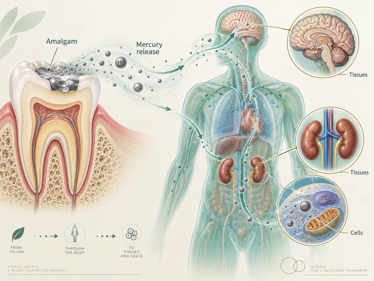
Where mercury vapor from amalgam fillings travels once inhaled, how it crosses into the brain and kidneys, why it stays for years, and what it does to cells.
Part 3 of 12 in the Mercury Series
Mercury Doesn’t Stay in the Tooth: Where It Goes and What It Does Once It Gets There
Part 3 of the Mercury Amalgam Series
In Part 1, we established that mercury doesn’t somehow become permanently trapped inside an amalgam filling just because the filling gets hard. In Part 2, we looked at what happens after some of that mercury leaves the restoration. Across multiple human studies, greater amalgam exposure generally meant more measurable inorganic mercury in blood or urine. So now we get to the question I REALLY care about:
Once mercury gets into the body, where does it go?
Because blood and urine only tell us part of the story. Blood tells us something about what’s circulating at a particular moment. Urine tells us something about what the body is getting rid of. Neither one gives us a complete inventory of what may already be sitting inside the brain, kidneys, thyroid, pituitary, or other tissues. And there’s an enormously important distinction buried inside that sentence:
Mercury leaving the body isn’t necessarily the same thing as mercury no longer being IN the body.
Before we go farther, let’s remind ourselves what we’re actually talking about. Dental amalgam is approximately 50% elemental mercury by weight, and even the FDA acknowledges that completed amalgam restorations release elemental mercury vapor that can be inhaled. [1]. Mercury is toxic. It’s a nonessential metal with no biological job that our bodies need it to perform. [A1]. The interesting questions aren’t whether mercury itself is toxic. We already know that.
The questions are how much gets into a particular person, where it goes, how long it stays there, what it does once it gets there, and why two people exposed to apparently similar amounts may handle it very differently. And THAT is where this story gets interesting.
A Quick Note About the Reference Numbers
Just like Parts 1 and 2, the regular reference numbers in brackets—such as [28] or [31]—are the exact same numbers used in the complete master mercury article. I’m keeping them consistent on purpose so you can move between these shorter articles and the master document without translating one bibliography into another. That means this article jumps from [1] to [28]. I haven’t lost references [2–27]. They support other parts of the mercury story.
You’ll also see references beginning with an A, like [A1]. Those are article-only references I added because while expanding this particular subject, I found additional science that deserved to be here. The monster master article is still the backbone. These shorter articles just give me room to follow an individual question further.
First, How Does Mercury Vapor Get From a Tooth Into the Rest of the Body?
This part matters because elemental mercury behaves VERY differently depending on whether you swallow it or breathe it. If somebody swallowed a little bead of liquid elemental mercury, very little would be absorbed through the gastrointestinal tract. But mercury vapor is a completely different exposure.
When elemental mercury becomes a vapor and reaches the lungs, it crosses that enormous lung surface area very efficiently. Reviews of elemental-mercury toxicology estimate that roughly 70–80% of inhaled mercury vapor can be absorbed into the bloodstream. [A1]. And amalgam releases vapor.
Once elemental mercury vapor enters the bloodstream, it can distribute throughout the body. Because elemental mercury—Hg⁰—is electrically neutral and lipid soluble, it can cross biological membranes relatively easily, including the blood-brain barrier. [A1]. Then something really important happens. Inside the body, elemental mercury can be oxidized into inorganic mercury—Hg²⁺.
That chemical change matters because inorganic mercury doesn’t cross lipid membranes nearly as easily as elemental mercury does. So elemental mercury can enter a tissue in one chemical form and then become a form that leaves that tissue much less easily. That helps explain why mercury vapor can enter the brain relatively efficiently while inorganic mercury may remain there for a VERY long time.
We’ve Actually Measured What Humans Retain After Breathing Mercury Vapor
We don’t have to build this entire explanation from animal research. There’s controlled human data. In one study, nine healthy volunteers who didn’t have amalgam fillings were exposed to a carefully measured concentration of elemental mercury vapor for 15 minutes. Researchers then repeatedly measured mercury in their blood, plasma, urine, and exhaled air for the next 30 days. [A4]
The median amount of inhaled mercury retained by the volunteers was approximately 69% of the dose. That number should get your attention. They inhaled mercury vapor, and roughly two-thirds of the inhaled dose was retained. During the first three days, only about 1% of the absorbed mercury was excreted in urine. By 30 days, the amount estimated to have been eliminated through urine varied enormously between individuals—from approximately 8% to 40% of the absorbed dose. [A4]
They got the same basic experimental exposure. They were different human beings. They experienced VERY different elimination. We aren’t cookie cutters.
And this is why a urine mercury measurement needs to be understood for what it actually is. It tells us something about mercury being excreted in urine. It doesn’t tell us where every mercury atom that ISN’T in the urine happens to be. Researchers also found substantial person-to-person variation in mercury concentrations in blood and plasma after the same exposure. [A4]. So when somebody takes one mercury value from one biological sample and treats it like a complete map of someone’s total body burden, I have a problem with that. You know what you measured. In that specimen. At that moment. That’s useful information. It is not a GPS tracker attached to every mercury atom in the body.
Human Autopsy Research Shows Mercury Beyond the Mouth
This is some of the evidence I find particularly compelling because researchers weren’t estimating tissue exposure from a blood or urine value. They actually analyzed human tissues. One autopsy study collected blood, brain, pituitary, thyroid, muscle, and toenail samples from 30 deceased adults. [28]. And importantly, they didn’t just measure “mercury” and leave us to guess where it came from.
They separated inorganic mercury from methylmercury.
That distinction matters because poor tuna gets dragged into practically every mercury conversation on Earth. . Seafood is primarily associated with methylmercury. Dental amalgam releases elemental mercury vapor, which is then converted largely into inorganic mercury inside the body. And the exposure patterns in this study reflected that difference. Methylmercury in the brain tracked with seafood exposure. But inorganic mercury in the brain was strongly associated with the number of dental amalgam surfaces present at death. Mercury in the pituitary and thyroid was also associated with amalgam burden. [28]
So the fish-associated mercury tracked with fish. The inorganic mercury tracked with amalgam. That makes it considerably harder to wave the finding away with: “Maybe they just ate a lot of tuna.”
And That Wasn’t the Only Human Autopsy Study
While expanding this article, I found more human tissue research that deserved to be included. Another autopsy study examined 34 people and measured mercury in several areas of the central nervous system as well as the kidney cortex. [A2]. Researchers found a statistically significant relationship between the number of tooth surfaces containing amalgam and mercury concentration in the occipital cortex of the brain.
And look at the kidney numbers! In the available kidney samples, seven people carrying amalgam had an average kidney-cortex mercury concentration of approximately 433 ng/g. Five people without amalgam averaged approximately 49 ng/g. [A2]. Those are small groups, but that’s almost a nine-fold difference.
And when researchers separated mercury forms in some of the tissue samples, an average of about 77% of the mercury was inorganic. [A2]. Another autopsy study examined 18 cadavers and compared people with more than 12 occlusal amalgam surfaces with people who had only 0–3. Mercury concentrations were significantly higher in the higher-amalgam group in the brain, thyroid, and kidney. [A3]
Again, these aren’t enormous studies. But these are actual human tissues. We now have multiple human autopsy studies associating greater amalgam burden with greater mercury accumulation in organs outside the mouth. So the sentence: “The mercury stays locked inside the filling.” simply doesn’t survive contact with the evidence.
Researchers Have Literally Traced Mercury From Fillings Through the Body
Animal research gives us an even cleaner way to answer the source question. Researchers made amalgam fillings using radioactively labeled mercury. [29]. Basically, they put a tracking device on the mercury. Then they placed those amalgams into monkey teeth and followed the labeled mercury through the body.
Within four weeks, mercury that had started specifically in those fillings had distributed into multiple organs and tissues. The highest concentrations were found in the kidneys, gastrointestinal tract, and jaw. [29]. This experiment answers a very simple question extremely well:
Can mercury that starts inside an amalgam filling leave that restoration and distribute elsewhere in the body? Yes. There’s no tuna to blame. No mystery occupational exposure. No childhood exposure we forgot to ask about. The researchers knew exactly where that mercury started.
It started in the fillings.
Getting Mercury Into a Tissue and Getting It Back OUT Are Two Completely Different Problems
This may be one of the most important concepts in the entire article. A blood mercury concentration going down doesn’t tell us that every tissue is now mercury-free. Neither does a falling urine concentration. Those measurements tell us what is circulating or being excreted in those particular compartments. A systematic review examined human poisoning cases, autopsy research, primate studies, and pharmacokinetic models to estimate how long inorganic mercury can remain in the brain. [30]
The answer was not a few days. Not even close. The evidence suggested that inorganic mercury can remain in the human brain for years and potentially decades. [30]. So imagine somebody had years of mercury exposure and then the exposure stopped. Their blood mercury may fall. Their urinary mercury may fall.
GOOD.
I want the exposure to stop and the biomarkers to come down. But if some mercury has already moved into a slowly exchanging tissue compartment, declining blood and urine values do not tell us exactly how much remains there.
Remember the controlled human vapor experiment? Thirty days after ONE defined exposure, estimated urinary elimination varied from approximately 8% to 40% of the absorbed dose between individuals. [A4]. That is why I think of urine primarily as an exit measurement. It tells us something about what is coming out. It isn’t a whole-body inventory.
Human biology simply isn’t cooperative enough to give us a convenient dashboard saying:
Brain: this much.
Thyroid: this much.
Kidneys: this much.
Pituitary: this much.
Everything else: all clear.
So WHY Does Mercury Matter Once It Gets Into Tissue?
Okay. We know mercury leaves amalgam. We know mercury vapor can enter the bloodstream. We know elemental mercury can cross biological membranes. We’ve measured amalgam-associated mercury in actual human brain, thyroid, pituitary, and kidney tissue. Now we get to the toxicology:
What does mercury actually DO once it gets there?
This is where the idea that mercury is somehow biologically inert really falls apart. Mercury is highly chemically reactive. It has a strong attraction to specific sites inside proteins and enzymes, and when it binds to those sites, it can interfere with the machinery our cells need in order to work.
Meet Thiols—And No, You Don’t Need to Memorize the Word
One major target involves sulfur-containing chemical groups called thiols. You don’t need to remember the word when you finish the article. There will not be a quiz. . But you do need to understand why they matter.
Thiols are found in molecules such as cysteine and glutathione and in many proteins throughout the body. [31]. Proteins aren’t random blobs floating around inside us. Their three-dimensional shape matters. Their binding sites matter. Their chemistry matters. They are tiny biological machines.
Mercury has a very strong attraction to sulfur-containing sites. When mercury binds to an important thiol group, it can change how that protein or enzyme functions. [31]. This affects systems involved in antioxidant defense, transport, metabolism, and cellular repair. And glutathione is part of that story.
Glutathione Is Important—but Mercury Toxicology Is Bigger Than One Supplement
Glutathione gets talked about constantly in mercury conversations. There’s a legitimate reason for that. Glutathione is one of the body’s major antioxidant and chemical-defense molecules, and its sulfur-containing groups participate in mercury binding, transport, and elimination. [31]
But mercury biology is considerably more complicated than: “Mercury affects glutathione, therefore take glutathione.” Glutathione is part of a larger network of redox systems that keep proteins functioning correctly, control reactive chemistry, and repair cellular damage. And mercury can interfere with several parts of that network.
Mercury Also Targets Selenium-Dependent Defense Systems
This is one of the pieces of additional science I wanted in the shorter article because it makes mercury toxicity even easier to understand. Selenium is incorporated into several enzymes that are essential to cellular redox control. One of them is thioredoxin reductase. Again, no quiz.
The important part is what the thioredoxin system does. It helps cells maintain proteins in the correct chemical state, manage oxidative stress, regulate growth, repair damage, and determine whether badly injured cells survive. [A5]. Mercury has an extremely high affinity for the selenium-containing active site of thioredoxin reductase.
Researchers have identified this enzyme as an important molecular target of mercury toxicity. Mercury can inhibit it and interfere with the cell’s ability to maintain normal redox chemistry. [A5]. This is why I get frustrated when mercury toxicology gets dismissed as vague “detox” language. We are talking about specific molecular targets and specific enzyme systems. That's biochemistry.
Oxidative Stress Is Not Just a Wellness Buzzword Either
Mercury research repeatedly brings us to oxidative stress. That phrase gets thrown around so much that I think people stop hearing it. So let’s make it simple.
Our cells naturally produce reactive molecules during normal metabolism.
That’s fine. We also have antioxidant systems that keep those reactive molecules under control. Oxidative stress happens when the production of reactive molecules exceeds the cell’s ability to control them. Then damage can begin to accumulate in: proteins, cell membranes, DNA, and mitochondria.
Mitochondria are the structures inside our cells that produce most of the energy the cells need. Researchers exposing kidney proximal-tubule cells to inorganic mercury found increased oxidative stress, abnormal intracellular calcium, disruption of the cell’s structural framework, and changes in autophagy—the process cells use to break down and recycle damaged components. [32]
Another study found that inorganic mercury disrupted normal mitochondrial dynamics and produced excessive oxidative stress in kidney cells. [33]. So now we have mercury interfering with:
-
sulfur-containing proteins,
-
glutathione-related chemistry,
-
selenium-dependent redox enzymes,
Account required
Sign in to see more
Create an account or sign in to unlock the full list.