Resource Libraries
Root Canal FAQ: Are Root Canals Safe? Informed Consent Guide
Answers to common root canal questions, why a root canal tooth can never be biologically equal to a living tooth, and dozens of peer-reviewed studies.
Part 1 of 2 in the Root Canal FAQ Series
FAQ's for Root Canals - Informed Consent, Biology, and Medical Freedom of Choice
There are certain questions that come up often enough in clinical practice and online education spaces that they deserve their own resource library article. Root canals are one of those topics, but they deserve multiple articles! So we decided to do just that. But as is customary for us, we fill our articles chock full of the SCIENCE, because the naysayers believe that biological dentistry operates on herbs and magic. Instead, we are SCIENCE FORWARD, so each article we write, we try to make sure that the science shines through. This article is no different. Be warned, there are over 30 PubMed articles sourced in this article alone. We have addressed the bacterial issue and the meridian interruption questions in separate articles already in our resource library. If you wish to read about those, here are the links:
Alternatives to Root Canals in Biological Dentistry:
Toxic Mold and Teeth: Why Root Canal Teeth Matter When Mold and Mycotoxins Won’t Clear https://www.orabiologics.com/resource-library/dental-relationships-to-toxic-mold%3A---why-root-canal-teeth-matter-when-mold-and-mycotoxins-won%E2%80%99t-clear
Oral Pathogens That Can Damage Your Whole Body: https://www.orabiologics.com/resource-library/oral-pathogens-in-root-canals-part-1
MTA and Root Canals:
https://www.orabiologics.com/resource-library/mta-and-root-canals
Root Canals in Permanent Front Teeth on Children:
https://www.orabiologics.com/resource-library/root-canals-on-permanent-front-teeth-in-children
People ask about materials. About techniques. About ozone. About regeneration. About scans that look “normal.” About a friend, a parent, or an influencer who seems to be doing just fine with a root canal decades later. And underneath all of those questions is usually the same unspoken one: Is it actually okay if I do this?
This article exists to answer some of the other frequently asked questions we get - honestly, without fear-mongering, without minimizing concerns, and without telling anyone what they should do with their body.
The Orabiologics and Biological Dentistry Philosophy (This Matters)
Let’s be very clear from the start:
-
We are not fans of root canal therapy. You will see why in ABUNDANT detail within this article alone, because in our honest opinion, the science does NOT support the validity of root canal therapy.
-
We are huge fans of informed consent and medical freedom of choice. So even if we don't love them, we will always support our patients if they choose this mode of dentistry as a treatment option.
Those two statements are not contradictory. Our role is not to persuade, pressure, or shame. Our role is to educate, to explain what the science shows (and does not show), to share what we see clinically over time, and to support people even when their choices differ from our personal preferences. Your body. Your choice. Always.
What a Root Canal Actually Is (Biologically)
A root canal removes the dying or dead and infected pulp tissue from inside a tooth and replaces it with supposedly inert materials in order to preserve the external structure of the tooth for chewing and appearance. I use that term "supposedly inert" because you will see the science shows it is NOT inert. These supposedly inert materials will be listed following this section, with all of the ingredients and the science associated with them. Once that pulp is removed, several things are permanently true:
-
There is no blood supply
-
No immune surveillance
-
No lymphatic drainage
-
No ability for the body to respond to infection from the inside
-
No ability to conduct bioelectrical energy cellularly
This is not controversial. It is basic anatomy. From that point forward, the tooth is no longer a living structure. It is a retained, non-vital object inside living bone, which acts like saran wrap on a plug. Everything else in this discussion flows from that reality.
Why bacteria can remain after a root canal (The part most people are never clearly told)
A root canal can be done carefully, skillfully, and exactly as taught — and it still cannot completely eliminate bacteria inside a tooth. This isn’t about poor dentistry. It’s about anatomy and biology.
Teeth are not simple hollow tubes
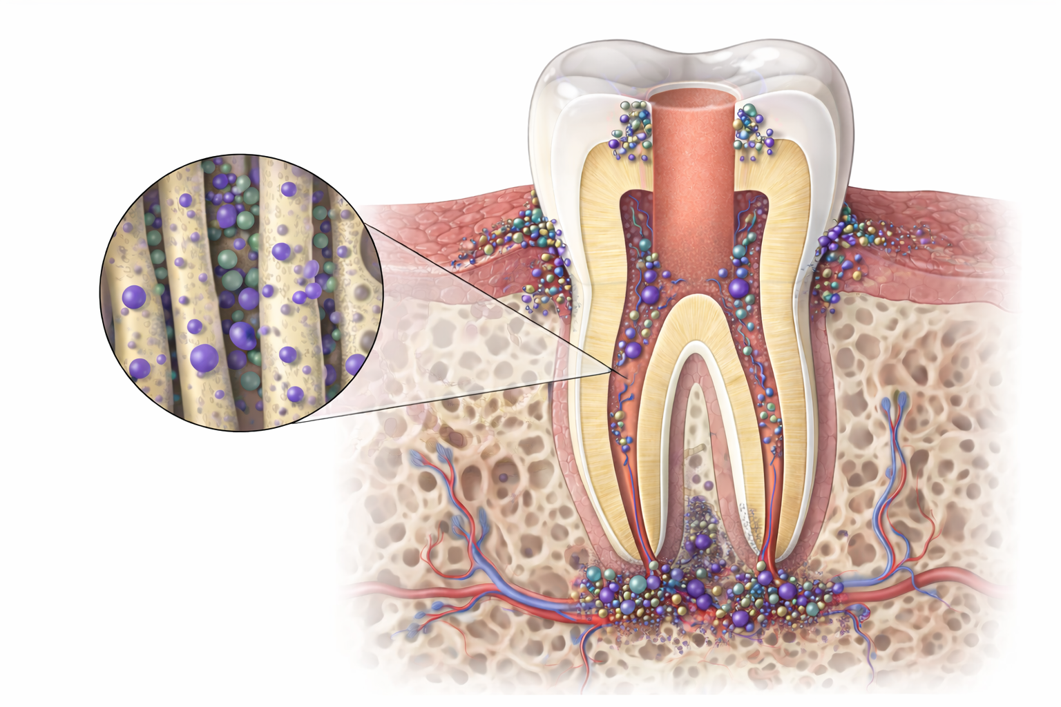
Inside a tooth, there isn’t just one open space. There is:
-
A main canal that dentists can access
-
Small side branches and connecting areas, especially near the root tip
-
And dentinal tubules — microscopic channels that extend outward from the canal into the tooth structure
When a tooth is alive, these channels carry fluid and cellular communication. After a root canal, the nerve and blood supply are gone — but the structure of these channels remains.
Dental instruments and disinfecting solutions can clean the main canal. They cannot reliably reach all of the side branches or penetrate deeply into the dentinal tubules.
What research consistently shows
When root-canal-treated teeth are examined under a microscope:
-
50–90% show ongoing inflammation or infection at the root tip
-
Many of these teeth look “successful” on X-rays and may not hurt
This tells us something important: A tooth can meet clinical success criteria and still not be biologically healed.
Why bacteria survive treatment
1. Some areas simply cannot be accessed. Bacteria can remain in:
-
Dentinal tubules beyond the cleaned canal
-
Small side canals
-
Branches near the tip of the root
These aren’t accidents — they’re part of normal tooth anatomy.
2. Bacteria behave differently inside teeth. Root canal infections are not made up of loose, floating bacteria. They exist as biofilms — organized communities attached to surfaces.
Biofilms:
-
Are harder to disinfect
-
Protect bacteria from chemicals
-
Allow bacteria to survive harsh conditions
This is why root canal infections behave very differently from infections in living tissue.
3. These infections are complex, not single-organism problems. Modern DNA testing shows:
-
Many different bacterial species can be present
-
Some cannot be grown or identified with traditional lab methods
-
Persistent infections are usually polymicrobial, not caused by one “bad” bacterium
The old idea that failure comes down to a single organism is outdated.
The sealing issue patients aren’t told about
Root canal treatment works by sealing the inside of a tooth. What it does not do is restore:
-
Blood flow
-
Immune cells
-
Detoxification
-
Repair mechanisms
The materials seal around whatever remains inside the tooth. There is no evidence that sealing bacteria inside a tooth makes them harmless long-term. In fact, studies show bacteria can attach to and grow on root canal filling materials themselves.
Even when bacteria are reduced, toxins remain
Bacteria leave behind inflammatory byproducts. These toxins:
-
Persist after treatment
-
Are found in failed root canals
-
Are associated with larger areas of bone inflammation around the tooth
So the issue isn’t only living bacteria — it’s the biological activity they leave behind.
The core limitation that changes everything
A root-canal-treated tooth is non-vital. That means:
-
No immune surveillance inside the tooth
-
No way to clear toxins
-
No internal repair
-
No biological buffering if materials degrade over time
Living tissue can heal. A dead structure can only be sealed and monitored. And root canals are the only thing that medicine seems to think is ok to leave in the body. If literally any other part of the body dies, we must do surgery to remove that body part. Gangrenous toe? Amputate it. Appendicitis? Appendectomy. But for some reason, dentistry seems to have missed that message and continues to insist that it's ok to keep something dead in the body if it's "taxidermied".
What the body appears to be doing
Chronic inflammation at the root tip may actually be a containment strategy — the body isolating a sealed but biologically active space to protect the rest of the system. In that sense, post-treatment inflammation isn’t the immune system failing. It may be the immune system doing its best with limited options. The problem is though that we have shown repeatedly in our resource library, which at this point is filled with PubMed research, that the situation never remains local. It simply buys TIME. Which you get to decide, as a patient, if that's ok and aligns with your values! Because it's ALWAYS going to be your body, your choice. Nobody should ever make you feel helpless in this situation. You are the one who gets to make the decisions about your body. Period. But in biological dentistry, we are going to give you the information WE have to help you make a TRULY INFORMED DECISION. Because we believe you have the right to make that decision. FULL STOP.
NEXT is where we discuss the SCIENCE. Because of course we have to pay attention to the science! Biological dentistry is SCIENCE FORWARD ALWAYS. That part is not negotiable for us!
What materials are used in a root canal?
A completed root canal typically involves four categories of materials:
1. Root canal filling material (inside the tooth)
Gutta-percha
This is the main material used to “fill” the cleaned canal space.
What it’s made of:
-
~20% gutta-percha polymer (a latex-like material from tropical trees)
-
~60–75% zinc oxide
-
Heavy metal salts for radiopacity (commonly barium sulfate)
-
Plasticizers, waxes, resins
-
Sometimes antimicrobial additives
Important nuance:
Despite the name, most of what’s inside the tooth is not gutta-percha — it’s zinc oxide plus additives.
Friedman CM, Sandrik JL, Heuer MA, Rapp GW.
PMID: 1058875
https://pubmed.ncbi.nlm.nih.gov/1058875/
Gutta-percha filling points contain about 20% gutta-percha polymer, 66% zinc oxide filler, 11% heavy metal sulfates for x-ray visibility, and 3% waxes or resins. The mechanical properties depend mainly on how much gutta-percha and zinc oxide are present.
-
Clinical Takeaway: This classic 1975 study established the standard composition of gutta-percha endodontic points. The high zinc oxide content (66%) is particularly relevant to our resource library theme, as this component has been identified in later studies as a nutrient source that supports fungal growth, especially Aspergillus species, when gutta-percha accidentally enters the maxillary sinus or remains in infected root canals.
Now here is where we refer to the issues with root canal materials, particularly zinc, feeding mold. In our toxic mold and teeth library, we listed several studies that showed these issues. Here is the link to that library resource, but following will be a dozen more studies that show the same issue:
Toxic Mold and Teeth: Why Root Canal Teeth Matter When Mold and Mycotoxins Won’t Clear https://www.orabiologics.com/resource-library/dental-relationships-to-toxic-mold%3A---why-root-canal-teeth-matter-when-mold-and-mycotoxins-won%E2%80%99t-clear
Here are additional studies, because it's important that you see that multiple studies exist on the subject, and it's not an outlier kind of situation:
1. Senges C, Wrbas KT, Altenburger M, et al.
PMID: 21846541
https://pubmed.ncbi.nlm.nih.gov/21846541/
This study tested how well bacteria and Candida fungus stick to different root canal filling materials. Gutta-percha and Real Seal SE had the highest bacterial adhesion, while Apexit Plus had less. All materials allowed microorganisms to attach and potentially form biofilms, which can lead to treatment failure.
-
Clinical Takeaway: Root canal filling materials, especially gutta-percha, provide surfaces where bacteria and fungi readily adhere and form mixed biofilms. The material itself becomes a substrate that supports persistent polymicrobial colonization even after treatment.
2. Neelakantan P, Romero M, Vera J, et al.
PMID: 28800075
https://pubmed.ncbi.nlm.nih.gov/28800075/
This review explains how bacteria form complex, organized communities called biofilms in root canals. These biofilms are fundamentally different from free-floating bacteria and are much harder to kill. The complexity of root canal anatomy combined with multi-species biofilms makes complete disinfection extremely challenging.
-
Clinical Takeaway: Biofilms in root canals represent organized microbial ecosystems where bacteria and fungi cooperate, share nutrients, and protect each other. Foreign materials in the canal system provide scaffolding for these communities, fundamentally altering the biology from planktonic to sessile growth.
3. Long J, Kreft JU, Camilleri J.
PMID: 32014453
https://pubmed.ncbi.nlm.nih.gov/32014453/
This study exposed different root canal materials to bacterial challenge for eight weeks. Gutta-percha and certain sealers encouraged thick biofilm accumulation, while some materials resisted biofilm formation. Importantly, bacterial biofilms caused structural changes in some materials, including washout and crystal deposition.
-
Clinical Takeaway: Dental materials not only support biofilm growth but are actively degraded by microbial metabolic byproducts. This bidirectional interaction—where materials feed pathogens and pathogens degrade materials—creates a self-perpetuating cycle of infection and material breakdown.
4. Siqueira JF, Rôças IN.
PMID: 34958494
https://pubmed.ncbi.nlm.nih.gov/34958494/
This comprehensive review discusses how bacteria form sessile communities (biofilms) attached to root canal walls. Over 500 bacterial species have been detected in endodontic infections, with 20-30 core species most common. Bacterial interactions within these communities determine overall virulence, making the community itself the unit of disease.
-
Clinical Takeaway: The pathogenicity of endodontic infections stems from community-level interactions rather than individual species. Foreign materials provide the physical substrate where these polymicrobial communities assemble, communicate, and express enhanced virulence through cooperative metabolism.
5. Al-Sakati H, Kowollik S, Gabris S, et al.
PMID: 32894604
https://pubmed.ncbi.nlm.nih.gov/32894604/
This study analyzed bacteria and fungi in removed gutta-percha from failed root canals. Molecular methods detected microorganisms that traditional culture missed, including difficult-to-grow bacteria. Candida DNA was found in gutta-percha samples, showing that filling materials themselves harbor mixed bacterial-fungal communities.
-
Clinical Takeaway: Gutta-percha and other filling materials actively harbor polymicrobial communities including fungi. These materials serve as reservoirs where bacteria and fungi coexist, with some organisms only detectable by molecular methods, indicating the materials support diverse, complex microbial ecosystems.
6. Chavez de Paz LE.
PMID: 17509401
https://pubmed.ncbi.nlm.nih.gov/17509401/
This review challenges the idea that persistent infections are caused by "super-resistant" bacteria. Instead, it proposes that bacteria adapt their physiology to survive treatment by forming biofilms. Surface adherence to materials triggers genetic changes that significantly increase bacterial survival, making the biofilm community more resistant than individual cells.
-
Clinical Takeaway: Dental materials trigger adaptive genetic responses in bacteria, transforming them from vulnerable planktonic cells to highly resistant biofilm phenotypes. This material-induced physiological shift represents a fundamental biological change where the substrate itself programs enhanced microbial survival.
7. Abusrewil S, Alshanta OA, Albashaireh K, et al.
PMID: 32233822
https://pubmed.ncbi.nlm.nih.gov/32233822/
This critical review examines endodontic biofilm infections and their polymicrobial nature. Despite advances in clinical techniques, treatment outcomes haven't improved significantly. The review emphasizes understanding biofilm polymicrobiality and host response to develop better diagnostic, treatment, and prevention strategies for endodontic disease.
-
Clinical Takeaway: Polymicrobial biofilms on dental materials represent a distinct pathogenic entity requiring different treatment approaches than planktonic infections. The material-biofilm interface creates a protected niche where cross-kingdom interactions between bacteria and fungi enhance overall community resistance.
8. George S, Basrani B, Kishen A.
PMID: 20630308
https://pubmed.ncbi.nlm.nih.gov/20630308/
This study showed that Enterococcus faecalis forms biofilms on gutta-percha under both nutrient-rich and nutrient-poor conditions. When gutta-percha was coated with saliva or serum proteins, bacteria adhered even better. This demonstrates that gutta-percha can support biomaterial-centered infections similar to other medical implants.
-
Clinical Takeaway: Gutta-percha conditioned with biological fluids (saliva, serum) becomes an enhanced substrate for bacterial colonization. Protein conditioning creates a biological interface that facilitates initial adhesion and subsequent biofilm formation, even under nutrient-deprived conditions that would normally inhibit growth.
9. Wang Z, Shen Y, Haapasalo M.
PMID: 34947188
https://pubmed.ncbi.nlm.nih.gov/34947188/
This review examines antimicrobial properties of bioceramic materials used in endodontics. Different materials showed varying abilities to resist biofilm formation. Some bioceramics prevent bacterial adhesion, while others allow thick biofilm accumulation. New supplements are being developed to enhance antibiofilm properties of these materials.
-
Clinical Takeaway: Material composition directly determines biofilm susceptibility. While some bioceramics resist microbial colonization through ion release and pH modulation, others provide favorable surfaces for mixed bacterial-fungal biofilm development, highlighting how material chemistry fundamentally shapes the microbial ecology.
10. Negrini TC, Koo H, Arthur RA.
PMID: 31732939
https://pubmed.ncbi.nlm.nih.gov/31732939/
This review discusses cross-kingdom interactions between Candida and bacteria in oral biofilms. Candida has been found in polymicrobial biofilms associated with root canal infections. These Candida-bacterial interactions are critical for co-existence and may affect disease severity, with each organism supporting the other's survival.
-
Clinical Takeaway: Candida-bacterial biofilms represent synergistic cross-kingdom communities where fungi and bacteria mutually enhance each other's virulence. Dental materials provide the substrate where these cooperative interactions occur, with fungal hyphae creating scaffolding for bacterial attachment and bacteria providing nutrients that support fungal growth.
11. Karygianni L, Anderson AC, Tennert C, et al.
PMID: 24677170
https://pubmed.ncbi.nlm.nih.gov/24677170/
This study sampled both root canals and the filling materials themselves from failed treatments. They found 22 different bacterial species, with 9 species detected only in the filling materials. This proves that obturation materials harbor distinct microbial communities separate from the canal walls.
-
Clinical Takeaway: Endodontic filling materials develop their own distinct microbiome separate from canal wall biofilms. These material-associated communities include species not found elsewhere in the canal, demonstrating that foreign materials create unique ecological niches that support specialized microbial populations.
12. Prada I, Micó-Muñoz P, Giner-Lluesma T, et al.
PMID: 31041915
https://pubmed.ncbi.nlm.nih.gov/31041915/
This review identifies microorganisms associated with endodontic failure and why they survive disinfection. Key survival mechanisms include biofilm formation, hiding in unreachable areas, synergistic relationships between species, expression of survival genes, and activation of alternative metabolic pathways that allow growth on unusual nutrient sources.
-
Clinical Takeaway: Persistent microorganisms survive by exploiting material surfaces for biofilm formation, utilizing synergistic metabolic cooperation, and activating stress-response genes. Dental materials provide both physical protection and alternative nutrient sources through material degradation, enabling metabolic flexibility that sustains mixed microbial communities.
13. Hao Y, Huang X, Zhou X, et al.
PMID: 30322190
https://pubmed.ncbi.nlm.nih.gov/30322190/
This review examines how dental material surfaces influence biofilm formation. Chemical composition (organic matrix, inorganic fillers, fluoride, metallic ions) can enhance or inhibit biofilm growth. Surface roughness, topography, free energy, and hydrophobicity all significantly affect bacterial colonization and biofilm development on materials.
-
Clinical Takeaway: Material surface properties—chemistry, topography, roughness, and energy—fundamentally determine microbial colonization patterns. Irregular surfaces and specific chemical compositions create favorable microenvironments for bacterial-fungal biofilm assembly, with metallic ions and organic matrices potentially serving as nutrient sources for polymicrobial communities.
14. Kreth J, Merritt J, Pfeifer CS, et al.
PMID: 32479134
https://pubmed.ncbi.nlm.nih.gov/32479134/
This review discusses how oral microbiomes interact with dental composite materials. Microbial metabolic activities directly degrade materials, contributing to restoration failure. The acquired salivary pellicle forms on material surfaces, providing attachment sites for initial colonizers, followed by integration of additional species into mature polymicrobial biofilms.
-
Clinical Takeaway: Dental materials undergo biological conditioning through salivary pellicle formation, creating attachment sites for pioneer species. Subsequent microbial succession builds diverse polymicrobial biofilms whose metabolic byproducts actively degrade the material, establishing a destructive feedback loop between material breakdown and enhanced microbial nutrition.
15. Nicolai P, Mensi M, Marsili F, et al.
PMID: 26019392 (PMC4443559)
https://pmc.ncbi.nlm.nih.gov/articles/PMC4443559/
This study measured zinc and other metals in fungal balls from sinuses and compared them to dental filling materials. Similar metal concentrations were found in fungus balls and root canal materials, suggesting that dental materials accidentally pushed into sinuses provide nutrients (especially zinc) that help fungus grow.
-
Clinical Takeaway: Zinc oxide and other metallic components in dental materials serve as direct nutrient sources for fungal growth, particularly Aspergillus species. This demonstrates how material composition doesn't just provide a surface for colonization but actively feeds pathogens through leaching of bioavailable metals.
16. Yoo YJ, Kim AR, Perinpanayagam H, et al.
PMID: 32858856
https://pubmed.ncbi.nlm.nih.gov/32858856/
This review explains how Candida fungus infects root canals and resists treatment. Candida adheres to tooth structure, forms protective biofilms, and penetrates into dentinal tubules where disinfecting solutions cannot reach. This makes fungal root canal infections extremely difficult to eliminate with standard treatments.
-
Clinical Takeaway: Candida exploits the three-dimensional architecture of root canal systems and dental materials by forming biofilms and penetrating tubular structures. These fungal biofilms create protective matrices that exclude antimicrobials while establishing metabolic cooperation with co-colonizing bacteria in mixed-species communities.
17. Terashima K, Kagoya R, Tanaka M, Nishijima H, Kondo K.
PMID: 39463579
https://pubmed.ncbi.nlm.nih.gov/39463579/
This case report describes a patient who developed both fungal sinus infection and bacterial infection after root canal filling material accidentally entered the sinus cavity. The dental material (gutta-percha) created an environment where both Candida fungus and bacteria could grow, causing sinus problems requiring surgery to remove.
-
The clinical takeaway: This case highlights something dentistry does not like to talk about:
foreign materials change biology. When root canal materials enter the sinus:
They act as a physical scaffold
They trap moisture
They support mixed bacterial–fungal biofilms
Systemic medications and detox protocols do not reliably reach these spaces. Surgical removal was required because the body had no other mechanism to resolve the infection.
These articles collectively demonstrate that dental materials are not biologically inert—they actively participate in creating and sustaining polymicrobial infections through multiple mechanisms: providing adhesion surfaces, releasing nutrients (especially metals like zinc), undergoing degradation that feeds microorganisms, and creating protected niches where bacterial-fungal biofilm communities develop enhanced resistance and virulence through cooperative metabolism.
2. Root canal sealers (the “glue”)
Gutta-percha cones do not seal by themselves. Sealers fill microscopic gaps and dentinal tubules.
Common sealer types:
a) Zinc oxide–eugenol (ZOE) sealers
-
Zinc oxide
-
Eugenol (clove oil derivative)
-
Resin modifiers
Concerns: Same exact concerns as the 16 resources in the Gutta Percha section above. I didn't see the point of repeating the same thing.
b) Epoxy resin–based sealers (e.g., AH Plus)
-
Bisphenol-A–related epoxy resins
-
Amines
-
Fillers
Concerns:
-
Release of formaldehyde during setting (low but measurable)
-
Persistent cytotoxicity
-
BPA-related chemistry
1. Huang TH, Yang JJ, Li H, Kao CT.
PMID: 11762857
https://pubmed.ncbi.nlm.nih.gov/11762857/
Both AH26 and AH Plus epoxy resin sealers caused dose-dependent toxic effects on brain cells and damaged DNA. The study showed these materials harm both cell survival and genetic material, raising concerns about their safety when used near living tissues.
-
Clinical Takeaway: This foundational study demonstrated that epoxy resin-based sealers exhibit both cytotoxicity and genotoxicity in vitro. The dose-dependent DNA damage suggests that material leachates—likely including bisphenol-A derivatives and amine hardeners—can cause genetic injury to periapical cells.
2. Athanassiadis B, George GA, Abbott PV, Wash LJ.
PMID: 25283681
https://pubmed.ncbi.nlm.nih.gov/25283681/
This review examines how formaldehyde is released from epoxy resin endodontic materials like AH 26 during the chemical setting reaction. The article discusses whether the amount released is enough to overwhelm the body's natural ability to handle formaldehyde and cause harm.
-
Clinical Takeaway: Formaldehyde release occurs during epoxy resin polymerization as a byproduct of the amine-epoxide reaction. While the body has endogenous formaldehyde metabolism pathways, the review questions whether localized concentrations from dental materials exceed physiological detoxification capacity in periapical tissues.
3. Lodienė G, Kopperud HM, Ørstavik D, Bruzell EM.
PMID: 24028598
https://pubmed.ncbi.nlm.nih.gov/24028598/
This study identified specific chemicals leaking from root canal sealers. The epoxy-based material released bisphenol-A diglycidyl ether (BADGE), while methacrylate materials released different monomers. Fresh materials caused more cell death than fully hardened ones, with effects varying by material type.
-
Clinical Takeaway: BADGE (bisphenol-A diglycidyl ether) is the primary leachable from epoxy-based sealers and represents the unreacted epoxy resin component. This bisphenol-A derivative demonstrates time-dependent cytotoxicity, with fresh material showing greater toxicity than cured specimens due to higher concentrations of unreacted monomers.
4. Malta CP, Barcelos RCS, Fernandes PS, et al.
PMID: 38353803
https://pubmed.ncbi.nlm.nih.gov/38353803/
Computer modeling predicted that bisphenol-A epoxy resin from AH Plus has the highest toxicity among tested compounds, including liver toxicity, cancer risk, and irritation. It also showed the strongest binding to inflammatory proteins, suggesting it may worsen inflammation around tooth roots.
-
Clinical Takeaway: In silico toxicology predictions identified bisphenol-A epoxy resin as having the lowest maximum tolerated dose and greatest number of toxicological endpoints (hepatotoxicity, carcinogenicity, irritancy). Molecular docking showed strong affinity for inflammatory cytokines (IL-1β, IL-6, IL-8, TNF-α), suggesting direct immunomodulatory effects.
5. Jung S, Sielker S, Hanisch MR, et al.
PMID: 29579090
https://pubmed.ncbi.nlm.nih.gov/29579090/
AH Plus was highly toxic to bone-forming cells when freshly mixed but became safe after hardening. In contrast, some other sealers remained toxic even after setting. This shows that fresh AH Plus should never touch bone cells during dental procedures.
-
Clinical Takeaway: Fresh AH Plus exhibits severe cytotoxicity to osteoblasts due to high concentrations of unreacted epoxy resin and amine components. Cytotoxicity resolves after complete polymerization, indicating that leachable monomers—not the polymerized matrix—drive the toxic response. Clinical technique must prevent fresh material contact with periapical tissues.
6. Troiano G, Perrone D, Dioguardi M, et al.
PMID: 29593163
https://pubmed.ncbi.nlm.nih.gov/29593163/
Three epoxy resin sealers (AH Plus, Sicura Seal, Top Seal) all showed moderate toxicity to bone cells both when directly touching cells and through released chemicals. After one week, direct toxicity disappeared but indirect toxicity from released substances remained.
-
Clinical Takeaway: All epoxy resin-based sealers demonstrated persistent indirect cytotoxicity even after one week, indicating continued leaching of toxic components. The sustained release of bisphenol-A derivatives and amine compounds creates ongoing exposure to periapical tissues beyond the initial setting period.
7. Camargo CH, Oliveira TR, Silva GO, et al.
PMID: 24666905
https://pubmed.ncbi.nlm.nih.gov/24666905/
Setting time significantly affected how toxic root canal sealers were to cells and DNA. AH Plus became less toxic to cells after 12 hours of hardening, but DNA damage from all sealers actually increased at 24 hours, showing that hardening time matters for safety.
-
Clinical Takeaway: While cytotoxicity decreases with polymerization, genotoxicity paradoxically increases at 24 hours for all sealers tested. This suggests that secondary degradation products or oxidative byproducts generated during the curing process may cause DNA damage distinct from the direct cytotoxic effects of unreacted monomers.
8. Al-Hiyasat AS, Tayyar M, Darmani H.
PMID: 20078704
https://pubmed.ncbi.nlm.nih.gov/20078704/
Among four resin-based sealers tested, AH Plus showed the lowest toxicity to cells, while methacrylate-based sealers were more toxic. Diluting the materials by ten times greatly reduced toxicity for most sealers, showing that concentration of released chemicals determines how harmful they are.
-
Clinical Takeaway: AH Plus demonstrated superior biocompatibility among resin-based sealers, with dose-dependent cytotoxicity that significantly decreased upon dilution. This suggests that while epoxy resin components are inherently toxic, the total leachable concentration from AH Plus is lower than methacrylate-based alternatives.
9. Wuersching SN, Diegritz C, Hickel R, Huth KC, Kollmuss M.
PMID: 35660956
Account required
Sign in to see more
Create an account or sign in to unlock the full list.