Resource Libraries
Is Mercury in Amalgam Fillings Dangerous? What Science Shows
Whether mercury in amalgam fillings is dangerous, how much vapor a filling releases each day, and why the removal method matters as much as the decision.
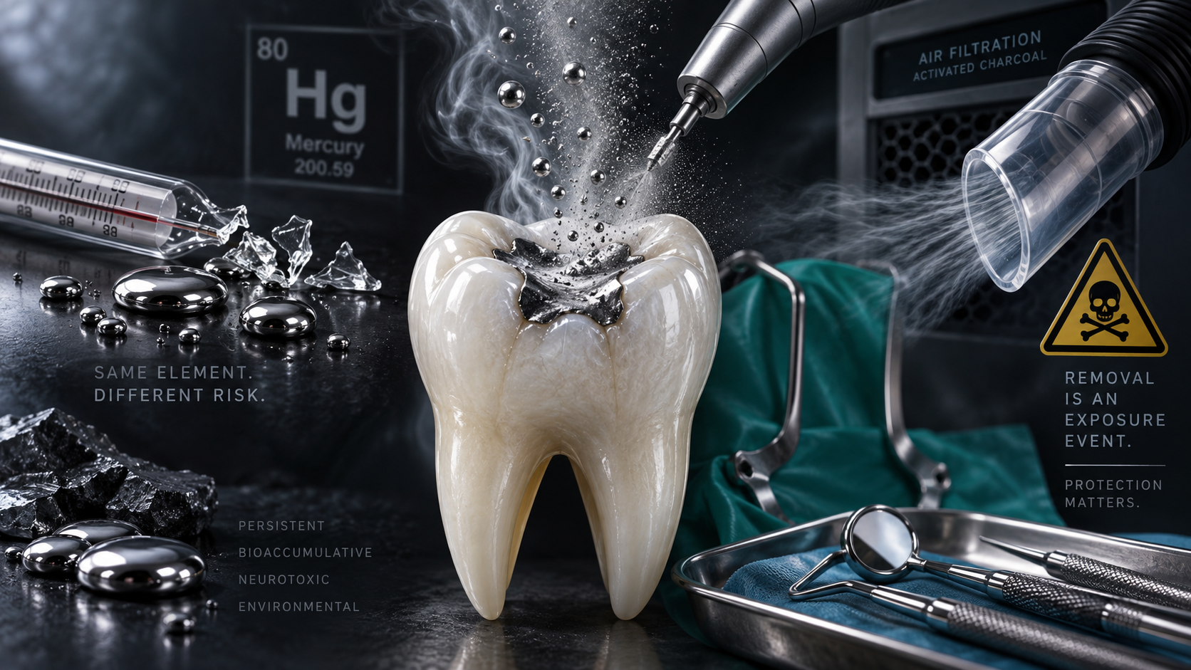
Mercury Amalgam Is Not an Inert Filling: What the Science Actually Shows and Why the Removal Method Matters
There’s a fundamental contradiction embedded in modern dentistry that most patients, and honestly quite a few dentists, have never been asked to confront. Dental amalgam is approximately 50% elemental mercury by weight. [1,2] While it’s sitting in a human tooth, patients are routinely told that it’s stable, inert, and perfectly safe. The moment that same filling is drilled out, however, the pieces, particles, suction-line residue, traps, and wastewater suddenly have to be collected and managed so that the mercury doesn’t contaminate the dental office, the sewer system, the environment, or the people handling it. [3]
This is where the contradiction gets even harder for me to understand. The EPA estimates that an average amalgam filling contains approximately 300 mg of mercury when it’s removed from a tooth. [3] An old-fashioned oral mercury thermometer contains approximately 610 mg. [4] In other words, roughly two average removed amalgam fillings contain about the same amount of mercury as one mercury fever thermometer. Now take that mercury out of somebody’s mouth and put it on the floor. Suddenly, everybody remembers that mercury is hazardous.
Even for a mercury spill approximately the size of the amount contained in a thermometer, EPA instructions say to get everyone else out of the area, remove pets, open windows and exterior doors, and close the room off from the rest of the building. You’re specifically told not to vacuum it, because a vacuum can put more mercury into the air and increase exposure. You’re told not to sweep it, because a broom breaks the mercury into smaller droplets and spreads it farther. After cleanup, EPA recommends continuing to ventilate the area to the outside for at least 24 hours, keeping children and pets away, and considering professional mercury-vapor monitoring to make sure residual vapor isn’t still present. [5] And that’s for approximately a thermometer’s worth or less.
If the spill is more than the amount contained in a thermometer, EPA instructions become even more serious. Everyone else is told to leave the area, exterior windows and doors are opened, doors to the rest of the building are closed, and the local health department or fire department is contacted for help. EPA also warns not to vacuum, sweep, pour the mercury down a drain, or walk through the contaminated area. [6] And cleanup isn’t considered finished simply because somebody can’t see little silver balls on the floor anymore.
After the visible mercury has been removed, EPA school guidance actually recommends temporarily heating the room to approximately 80–90°F while continuing to ventilate it to the outside. Why? Because heat increases mercury volatilization. The goal is to drive residual elemental mercury that may still be hiding in cracks, pores, or contaminated surfaces into the air so that ventilation can carry that vapor out of the building. EPA documented this same principle during an actual school mercury cleanup in Wyoming, where contaminated objects were taken to a heated area specifically to drive off mercury vapor. Afterward, the area is monitored for mercury vapor using a mercury-vapor analyzer or laboratory air sampling before people are allowed to reoccupy the room. [7,9]
The ventilation piece matters just as much as the heat. Mercury that has vaporized doesn’t politely stay in the room where the spill happened. If that contaminated air is pulled into the building’s heating and air-conditioning system, the HVAC system can carry that mercury vapor through the ductwork and redistribute it into other rooms. That’s why EPA specifically says to close the cold-air returns, turn off the ventilation and cooling systems serving the contaminated room, temporarily seal the air ducts if possible, and use fans only when they’re blowing the contaminated air outside. [7] So the goal isn’t merely to make the mercury disappear from sight. It’s to keep the vapor from being redistributed through the building, deliberately move any residual mercury into the air where it can be exhausted outdoors, and then measure the air afterward to make sure the remaining mercury vapor has fallen to an acceptable level.
OSHA workplace guidance is equally clear that mercury contamination isn’t just a “pick up what you can see and move along” problem. OSHA states that people who aren’t wearing appropriate respiratory protection should be excluded from areas where mercury spills or leaks have occurred until cleanup is complete. Its guidance also addresses contaminated clothing, footwear, work surfaces, respiratory protection, and methods of cleaning mercury-contaminated areas. [8]
So think about the absurdity of this for a minute. If mercury is dropped on the floor? Get people away from it. Isolate the room. Ventilate the building. Don’t vacuum it. Don’t sweep it. Protect the people cleaning it up. Keep contaminated clothing from carrying it elsewhere. Dispose of the waste appropriately. Heat the room afterward specifically to drive out residual mercury vapor. Then measure the air to make sure the mercury vapor has actually come down to an acceptable level.
I want to be very clear here because this distinction gets used all the time to make the mercury in amalgam sound like some entirely different substance from the mercury we’ve been talking about. It isn’t. The mercury used to make dental amalgam is liquid elemental mercury. It’s mixed with a powdered alloy made primarily of silver, tin, and copper because the mercury reacts with those metal particles and creates a material that can be packed and shaped into the hole left after decay is removed. Once it sets, the amalgam becomes hard enough to function as a dental restoration. [1] But the mercury didn’t disappear. It didn’t stop being mercury. And it most certainly didn’t stop getting out.
Even the FDA acknowledges that set amalgam restorations release elemental mercury in the form of vapor that can be inhaled and absorbed through the lungs. The FDA also specifically acknowledges that the amount of vapor released can increase with activities such as chewing and teeth grinding. [1] But we don’t have to stop with the FDA’s acknowledgement, because researchers have actually measured what happens to mercury vapor when people use their teeth normally.
In one human study, researchers continuously measured mercury evaporation from the mouths of people with amalgam fillings. Chewing gum caused mercury release to rise substantially, reaching as high as 2.7 ng/s after five minutes of chewing. Then the researchers did something particularly useful: they covered the amalgam teeth with individual plastic covers. Mercury evaporation immediately dropped by 89–100%. [10] That’s about as close as experimental science gets to pointing at the fillings and saying, “Yep. Right there.”
Another human study compared people with amalgam restorations with people who didn’t have any. Before chewing, the people with amalgam already had intraoral mercury-vapor concentrations approximately 9 times higher than the controls. After 10 minutes of chewing, mercury vapor in the amalgam group increased roughly six-fold over its own baseline, reaching approximately 54 times the concentrations measured in people without amalgam fillings. The amount released also increased with the number and type of amalgam restorations present. [11]
Chewing isn’t the only ordinary thing that increases mercury release. Brushing does it too. In a laboratory study that tested different amalgam surfaces under controlled conditions, brushing the restorations with toothpaste caused an immediate increase in mercury vapor release. The same study found that higher temperatures also increased mercury vapor coming off the amalgam. [12] Human measurements show the same heat effect. In the Björkman and Lind study, increasing rinse-water temperature from 35–45°C increased mean mercury evaporation by about 1.7 times. [10] So when we talk about heat increasing mercury release, we’re not talking about some abstract chemistry experiment that has nothing to do with normal life. People drink hot coffee. They drink hot tea. They eat hot food. Their mouths change temperature all day long.
Bruxism—grinding, clenching, and gritting—matters too, although the science here is more nuanced. A human study specifically examining nighttime tooth grinding found that bruxism may increase mercury uptake, but the measured effect was modest and smaller than the effect associated with chewing gum. [13] That doesn’t make the finding irrelevant. I personally question how much of a difference there might be between true grinders and people who primarily clench or grit their teeth, because those aren’t mechanically identical behaviors. I don’t know whether that distinction was adequately captured in this study, and I can honestly say that.
So for now, I think the right approach is simply to say exactly what the study found instead of turning it into something bigger than it was. The broader point is that mechanical wear and heat can change mercury release from amalgam, which makes the claim that mercury is simply locked away inside the filling very difficult to defend.
And Then There’s Polishing
Polishing deserves its own discussion because this is routinely done in dental offices without anyone in the room treating it as a mercury-exposure event. Most patients probably assume polishing an old amalgam is completely different from placing or removing one. The mercury measurements say otherwise. In one study, researchers measured the total mercury vapor generated during common dental procedures involving amalgam. Placement of a new amalgam restoration released approximately 6–8 μg of mercury vapor. Wet polishing released approximately 2–4 μg. Removal under water spray and high-volume evacuation generated approximately 15–20 μg.
But dry polishing released 44 μg. [14] Forty-four. In that experiment, dry polishing released roughly 5–7 times as much mercury vapor as placing the filling in the first place. [14] And remember what we just learned about heat and mechanical abrasion. Polishing creates friction. Friction creates heat. Heat increases mercury volatilization. Suddenly that number makes a whole lot more sense.
A separate clinical study measured 242 air samples directly from patients’ mouths during the setting, removal, and polishing of amalgam restorations. Mercury vapor was measured during all three procedures, with mean concentrations ranging from approximately 85–326 μg/m³. The larger the amalgam restoration being worked on, the higher the mercury-vapor concentration tended to be. [15]
So polishing an amalgam isn’t simply making an old silver filling nice and shiny. You are mechanically abrading a mercury-containing material, generating heat in the process, and measurably increasing the amount of mercury vapor present in the patient’s mouth. That doesn’t mean nobody can ever polish an amalgam (though to be quite honest, I would definitely tell people it's probably a good idea to consider not doing that...). We need to stop pretending the procedure is biologically irrelevant.
And if you’re wondering whether that burst of mercury during polishing just disappears into the room and never shows up in the patient, another study gives us a pretty uncomfortable answer. Researchers measured 24-hour urinary mercury in 43 patients before and after three different procedures: removing old amalgams, placing new amalgams, or polishing existing amalgams. Before treatment, the average amount of mercury excreted in the urine was 18.5 μg over 24 hours. One day after treatment, it rose to 56.3 μg after removal, 45.9 μg after placement, and 56.25 μg after polishing. [16]
Yes. You read that correctly. In this study, polishing produced essentially the same 24-hour urinary mercury level as removing the amalgam altogether, and the measured level after polishing was actually higher than after placement. Mercury excretion remained significantly elevated above the patients’ baseline throughout the eight-day follow-up, although all of the measured values remained below the World Health Organization urinary thresholds used by the researchers. [16] That last part is true, but I’m not going to leave it out simply because it complicates the story.
The study does not prove that polishing an amalgam causes clinical mercury poisoning. What it does prove is that polishing isn’t merely a cosmetic event happening on the surface of a filling. It can create enough additional mercury exposure that a measurable increase shows up in the patient’s urine afterward. That’s a very different statement from “the mercury is permanently locked inside the filling,” because the latter is exactly what the conventional world has taught for decades: once the amalgam gets hard, the mercury is supposedly locked inside the restoration and inert.
It isn’t.
And this is where individual susceptibility matters to me. A laboratory threshold or a population average can’t tell me exactly how much additional exposure one particular human being can tolerate at one particular moment. Someone whose health is already being challenged by multiple exposures, impaired clearance, illness, nutritional deficiencies, genetics, or other physiologic stressors may have far less room for another insult than someone who is otherwise thriving. I’m not claiming this study proves that one polishing appointment will push that person over the edge. It doesn’t. I’m saying that when an entirely avoidable dental procedure can measurably increase mercury exposure, I don’t think it makes sense to pretend the patient’s starting point doesn’t matter.
The next question is whether all of this mercury release actually translates into a measurable difference in the body over time. And again, we don’t have to guess. In the New England Children’s Amalgam Trial, researchers followed children with amalgam restorations and measured urinary mercury during years three through five. Every additional amalgam surface present was associated with an approximately 9% increase in current urinary mercury, while each additional posterior chewing-surface year of amalgam exposure was associated with about a 3% increase in cumulative urinary mercury excretion. [17] A separate analysis of the Casa Pia Children’s Amalgam Trial found the same basic pattern. As cumulative exposure to amalgam increased, urinary mercury increased with it. Children with average amalgam exposure had urinary mercury levels approximately 18–52% higher, depending on age, than children with no amalgam exposure. [18]
And this isn’t limited to those two clinical trials. A longitudinal Korean study followed hundreds of children over several years and found that both current amalgam exposure and accumulated amalgam exposure predicted urinary mercury concentrations. Different children, different country, different investigators, same basic relationship: the more amalgam exposure a person has, the more mercury we tend to measure leaving the body in the urine. [19]
The numbers become even harder to wave away when we look at large U.S. population data. A 2021 analysis used National Health and Nutrition Examination Survey—NHANES—data from 14,181 people across multiple age groups. That distinction matters because, despite the title of the paper focusing on children, the full analysis wasn’t limited to 14,181 children. [20] The researchers also tried to separate mercury coming from amalgam from mercury coming from seafood. Seafood consumption was associated with higher methylmercury, which is exactly what we would expect. But amalgam exposure was strongly associated with inorganic mercury in blood and with mercury in urine. Among participants who reported no fish consumption, people with more than five amalgam-restored surfaces had 103% higher total blood mercury, 221% higher blood inorganic mercury, and 221% higher creatinine-corrected urinary mercury than people without amalgam-restored surfaces. [20]
The age breakdown is difficult to ignore, but when I dug into the methods, I found an important detail. The abstract describes the youngest group as children “under six,” but the actual methods grouped participants into ages 1–6, 7–12, 13–21, 22–45, 46–65, and over 65. [20] That matters because this was an analytical category, not some magical biological cutoff. The study does not tell us that a child suddenly becomes less susceptible to mercury when they move from one age category into the next.
There’s another important detail hiding in the wording. The paper repeatedly refers to “amalgam restorations,” which makes it sound as though researchers counted whole fillings. But the underlying NHANES dental examination recorded restorations surface by surface—lingual, occlusal, facial/buccal, mesial, and distal—and the study’s own methods describe the exposure variable in terms of restored dental surfaces. [20,22] So “more than five amalgams” is better understood as more than five amalgam-restored tooth surfaces, not necessarily more than five separate fillings. In fact, one large MODBL amalgam on a single tooth can represent five surfaces all by itself.
And that raises another limitation that matters enormously to me as a dentist: surface count is not the same thing as mercury dose. NHANES recorded which surfaces contained amalgam, but it did not measure the physical volume of each restoration, its depth, the exact alloy formulation, or the actual mass of elemental mercury sitting in that individual tooth. [20,22] A tiny one-surface amalgam barely larger than a pencil tip and a huge MODBL restoration replacing most of the clinical crown are clearly not equivalent amounts of material, even though the database can only describe them using surface codes rather than an actual mercury mass.
In the youngest age category, ages 1–6, 43 participants had more than five amalgam-restored surfaces, and that group had the highest blood inorganic-mercury and urinary-mercury concentrations of any age category examined. [20] That is a striking finding, but it is still a group-level finding from a relatively small subgroup. It does not establish a magic “safe age,” and it cannot tell us the exact dose or susceptibility of one individual child. What it does tell us is that more amalgam-restored surfaces were associated with more measurable inorganic mercury—and that the youngest children in this dataset deserve far more attention, not less.
More recent NHANES research found the same basic relationship in adults. An analysis of adults ages 18–70 found significantly higher total and inorganic blood-mercury concentrations in people with amalgam compared with people without it. The number of amalgam surfaces also significantly correlated with blood inorganic mercury. Once again, more amalgam meant more measurable inorganic mercury in the body. [21]
Once researchers established that more amalgam surfaces generally mean more measurable mercury in the body, another question followed: how do those estimated exposures compare with health-based mercury-vapor reference values? A 2011 U.S. population model estimated daily mercury exposure from amalgam using national data on body weight, restored tooth surfaces, and the relationship between amalgam burden and urinary mercury. Even using the study’s least conservative amalgam scenario, the authors estimated that approximately 67.2 million Americans would exceed the dose associated with the U.S. EPA reference concentration and approximately 122.3 million would exceed the dose associated with California’s more protective reference concentration. [23]
A newer analysis using 2015–2018 NHANES data reached a similarly uncomfortable conclusion in adults. The researchers estimated that approximately 16.4 million adults, or 10.4%, received amalgam-derived mercury-vapor doses above the U.S. EPA-based limit they used, while approximately 85.9 million, or 54.3%, exceeded the more restrictive California EPA-based limit. The enormous difference between those numbers is a really good illustration of why the phrase “below the safety limit” doesn’t mean much unless somebody tells you which safety limit they’re talking about. [24] But there’s an enormous limitation to all of these models that matters to me just as much as the choice of safety threshold: they cannot tell us what happens inside one particular human being.
A model can plug in things like age, body weight, number of amalgam surfaces, and average relationships between amalgam burden and urinary mercury. What it cannot tell me is how much mercury one particular person will absorb, how much they will retain, where it will distribute in their tissues, how efficiently their kidneys will clear it, or how their genetics, nutrition, health status, cumulative exposures, or physiologic reserve will affect their response. And we already know that this variability is real. One published report described three people whose mercury uptake from their own amalgam fillings was dramatically higher than expected. Their urinary mercury ranged from 23–60 μg/day, their blood mercury was approximately 5–10 times the population average, and investigators could not identify another meaningful mercury source. After their amalgams were removed, their mercury levels returned toward normal. The authors’ point was not that everyone with amalgam absorbs that much mercury. The point was almost the opposite: People with apparently similar external exposures can have very different internal doses. [26]
Genetics can influence that variability too. In another human study, researchers found that genetic variants involved in glutathione production and mercury handling were associated with significant differences in mercury concentrations measured in blood, plasma, and urine. [27] So two people can encounter the same form of mercury and still not necessarily absorb, retain, process, or eliminate it the same way.
And this is where my philosophy is different from an approach that relies too heavily on what happens to the average person. Population data are useful. Averages are useful. Models are useful. They help us see patterns across thousands or millions of people. But the patient sitting in front of me is not a population average. My proposed approach is to protect the most susceptible person we reasonably can. If I design my precautions around the person with the least physiologic margin for error—the child, the pregnant woman, the person with impaired clearance, the genetically susceptible patient, the person whose body is already carrying multiple other stressors—then the person with greater physiologic reserve is protected too.
The reverse is not necessarily true. A safety approach built around what appears tolerable for most people cannot guarantee protection for the individual sitting at the vulnerable end of the spectrum. And we often have no reliable way to know ahead of time exactly who that person is. That matters enormously to me because mercury exposure from dentistry is not something we are powerless to change. We can choose not to place mercury-containing restorations. We can decide not to polish them. We can use appropriate protection when we remove them. When an exposure is avoidable, I don’t see the wisdom in asking, “How much can the average person tolerate?” before we act. I would rather ask, “How do we protect the person who can tolerate the least?” Because when we protect the most vulnerable person in the room, we don’t make everybody else less safe. We protect everybody else by default.
That’s also why I’m careful about how I describe these modeled exposure numbers. They are estimates, not blood tests, tissue measurements, diagnoses, or proof that 16 million—or 86 million—Americans have mercury poisoning. But the reverse is equally important: a population model also cannot prove that a particular individual is safe simply because their estimated exposure falls below a population-derived benchmark.
Pregnant women have been modeled separately as well. A 2024 NHANES analysis estimated that about 28% of pregnant women received amalgam-derived mercury-vapor doses above the U.S. EPA-based value used in the study and about 36% exceeded the California EPA-based value. But there’s an important limitation here too: although NHANES weighting represented approximately 1.67 million pregnant women nationally, the actual unweighted sample contained only 37 pregnant participants. [25] That makes the finding worth paying attention to. It does not make 37 women a crystal ball for every pregnant woman in America. And once again, that brings me right back to the same problem: Population estimates can tell us what may be happening across a population. They cannot tell us exactly what is happening inside the individual human being sitting in the dental chair. And we aren’t cookie cutters.
Mercury Doesn’t Stay Politely in the Mouth
So far, we’ve established that amalgam releases mercury, that ordinary activities can increase that release, and that greater amalgam exposure generally means more measurable mercury in blood and urine. But blood and urine are only part of the story. The next question is the one I really care about: once mercury gets into the body, where does it go?
Human autopsy research gives us a pretty direct answer. In one study, researchers collected blood, brain, pituitary, thyroid, muscle, and toenail samples from 30 deceased adults. They chemically separated inorganic mercury from methylmercury so they could distinguish mercury associated with seafood from the form more closely associated with elemental-mercury exposure. The number of amalgam surfaces present at death was strongly associated with inorganic mercury in the brain, as well as mercury in the pituitary and thyroid. [28] That distinction matters enormously. Seafood consumption was associated with methylmercury in the brain, while amalgam burden was associated with inorganic mercury. In other words, the researchers weren’t simply finding “mercury somewhere in the body” and assuming it came from dental fillings. They measured different forms of mercury and found different exposure patterns. [28]
Animal tracer studies help us understand how that movement can happen. Researchers made amalgam fillings using a radioactive mercury tracer and placed them in monkey teeth so they could literally follow mercury originating in the restorations. Within four weeks, mercury from those fillings had distributed into multiple organs and tissues, with the highest concentrations found in the kidneys, gastrointestinal tract, and jaw. [29] Obviously, monkeys are not humans, and an animal study cannot tell us what clinical disease an amalgam will cause in a person. What it can show us is that mercury released from a dental restoration does not necessarily remain confined to that tooth.
And once inorganic mercury reaches certain tissues, it may not leave quickly. A systematic review examining human cases, primate research, autopsy findings, and pharmacokinetic models concluded that inorganic mercury can remain in the human brain for years and potentially decades. [30] That matters because a blood or urine measurement tells us something about exposure and elimination at a particular point in time. It does not necessarily tell us how much mercury may already be retained in tissues.
What Does Mercury Actually Do Once It Gets There?
Knowing that mercury can reach human tissues is one thing. The obvious next question is: why do we care once it gets there? Mercury is chemically reactive. It has a strong attraction to sulfur-containing groups called thiols, which are found in proteins and in molecules such as cysteine and glutathione. Those sulfur groups help proteins maintain their shape and perform their jobs, and glutathione is one of the body’s major antioxidant and detoxification systems. When mercury binds to those sites, it can interfere with normal cellular chemistry. [31] That does not mean every mercury atom that enters the body automatically creates a disease. Biology is far more complicated than that. It means we understand several plausible and measurable ways mercury can interfere with normal cellular function, especially when enough mercury reaches a susceptible tissue or when the person has less ability to compensate for the exposure.
Researchers studying inorganic mercury in kidney cells have documented increased oxidative stress, disruption of the cell’s internal structural framework, abnormal increases in intracellular calcium, and changes in autophagy—the process cells use to break down and recycle damaged components. [32] Another laboratory study found that inorganic mercury disrupted the normal behavior of mitochondria and produced excessive oxidative stress in kidney cells. [33]
If “oxidative stress” sounds like one of those phrases everybody throws around without explaining, here’s what it actually means. Our cells constantly produce reactive molecules as part of normal metabolism, and antioxidant systems normally keep them under control. Oxidative stress happens when the production of those reactive molecules overwhelms the cell’s ability to neutralize them. At that point, they can begin damaging proteins, cell membranes, DNA, and mitochondria—the tiny structures that produce most of the energy our cells need.
So when we say mercury can create oxidative stress or interfere with glutathione, we’re not using vague wellness language. We’re talking about specific biochemical systems that researchers can measure inside cells. And that helps explain why organs responsible for processing and eliminating inorganic mercury—especially the kidneys—keep showing up in the toxicology literature.
The Kidneys Aren’t a Footnote
The kidneys deserve their own discussion because they’re one of the major organs responsible for handling and eliminating inorganic mercury. And this is another place where I think we have to look at all of the evidence, including the findings that are reassuring. The original New England Children’s Amalgam Trial randomized 534 children to receive either amalgam or mercury-free composite restorations and followed them for five years. At the end of the trial, the researchers found no statistically significant difference in the primary renal outcome, urinary albumin, between the two groups. The amalgam group did have significantly higher urinary mercury, but the main kidney measurement used in the original trial was reassuring. [34]
Then researchers went back and looked at the kidney data in considerably more detail. Average levels of the kidney biomarkers still weren’t significantly different between the groups. But something else appeared: during years three through five, children in the amalgam group were 1.8 times as likely to have microalbuminuria, meaning an abnormal amount of the protein albumin leaking into the urine. Ten children in the amalgam group had microalbuminuria at both the three- and five-year measurements, compared with only two children in the composite group. [35]
Why does that matter? Healthy kidneys are supposed to keep most albumin in the bloodstream. When more of it begins leaking into the urine, it can be an early sign that the kidney’s filtering system is under stress. But I want to be just as clear about the limitation: the researchers themselves said this finding might have occurred by chance, particularly because they examined multiple kidney outcomes, and they recommended that it be studied further. [35]
And importantly, another major randomized children’s trial did not reproduce that finding. The Casa Pia trial followed 507 children for seven years and examined several markers of kidney integrity, including albumin and markers of the kidney tubules. The researchers found no significant difference between the amalgam and composite groups in those renal markers and no significant increase in microalbuminuria with amalgam treatment. [36]
So if someone tells you the randomized trials proved that amalgam damages every child’s kidneys, that simply isn’t what the trials showed. But if someone tells you the kidney question was completely settled and nothing concerning ever appeared, that isn’t accurate either. One major trial was reassuring on its primary renal outcome, a later analysis of that same trial found a statistically significant microalbuminuria signal, and the second major trial did not reproduce it. That is exactly the kind of mixed evidence that deserves more investigation, not less.
The kidney story doesn’t end with those two randomized trials. Other human studies have found exactly the kind of early renal signals we would expect if inorganic mercury were stressing the kidney, particularly the proximal tubules. In one study of 101 healthy adults, people with amalgam had significantly higher blood and urinary mercury than people without amalgam. They also had significantly higher urinary NAG, gamma-glutamyltransferase, and albumin—all markers used to look for early renal stress or injury. Within the amalgam group, urinary NAG and albumin increased with the number of amalgam fillings, and albuminuria correlated with both blood and urinary mercury. [37]
A later study of 182 children found another renal signal. Urinary NAG, a marker of proximal-tubule injury, was significantly associated both with the presence of amalgam restorations and with urinary mercury. The relationship between urinary mercury and NAG followed a dose-effect pattern, and NAG was also associated with a marker of oxidative stress. The authors concluded that low-level mercury exposure from amalgam appeared to affect kidney tubular function, although they were appropriately clear that the finding needed additional confirmation. [38]
Even the Casa Pia data aren’t quite as simple as “the kidney results were negative.” A later reanalysis of those data reported a statistically significant dose-dependent relationship between cumulative amalgam exposure and urinary GST-alpha, another marker associated with proximal-tubule integrity. The estimated increase was about 10% over eight years at average amalgam exposure. [39] But this finding was formally challenged by the original Casa Pia investigators, who argued that the reanalysis did not preserve the randomized design properly and that a small increase in GST-alpha could reflect the kidney responding to mercury exposure without necessarily representing clinically meaningful kidney injury. [40] I think both sides of that argument belong here.
And there are negative studies too. A study of 403 children in Shanghai found slightly higher urinary mercury among children with amalgam but no significant difference in NAG or microalbumin. [41] That matters. If I’m going to argue that there’s a renal signal, I also have to acknowledge when investigators looked for that signal and didn’t find it.
When researchers pulled the randomized evidence together in a 2020 systematic review and meta-analysis, they did not find statistically significant evidence that amalgam caused nephrotoxicity compared with composite. But the review also found that the evidence base was surprisingly thin: only six publications met the inclusion criteria, five were judged to have a high risk of bias, and only two studies could actually be pooled in the meta-analysis. Trial-sequential analysis concluded that the available evidence was underpowered because the required information size had not been reached. [42]
That distinction matters to me. “We do not currently have enough evidence to prove clinically significant nephrotoxicity” is not the same statement as “we have proven there is no renal effect.” What I see instead is a pattern: mercury is a known renal toxicant; the kidney accumulates inorganic mercury; several human amalgam studies have reported increases in early renal biomarkers such as albumin, NAG, gamma-GT, or GST-alpha; other studies have not reproduced those findings; and the randomized evidence remains too limited to settle the question conclusively. That's not a slam dunk for kidney disease. But it is more than enough for me to say there is a renal signal worth taking seriously, especially when the exposure itself is avoidable.
The FDA’s own recommendations make more sense in that context. The agency specifically lists people with impaired kidney function among the groups who may be more susceptible to mercury from dental amalgam and recommends avoiding new amalgam placement in these patients when possible and appropriate. The FDA also acknowledges that mercury localizes in tissues including the kidneys and that very limited clinical information exists about the effects of amalgam-derived mercury vapor in people whose kidneys are already impaired. [1]
To me, that’s the practical point. We may not have a randomized trial proving that amalgam causes chronic kidney disease. But we do have a known renal toxicant, measurable kidney accumulation, several human studies showing early renal biomarker signals, incomplete and underpowered randomized evidence, and an FDA warning specifically identifying impaired kidney function as a higher-risk condition. I don’t need to pretend the evidence is stronger than it is to decide that adding more mercury to someone whose kidneys are already struggling makes absolutely no sense to me.
Pregnancy: The Placenta Isn’t a Force Field
Pregnancy raises the stakes because now we aren’t only asking what happens inside one adult body. We’re asking what happens when that mercury reaches the placenta and the developing fetus. And the placenta does not simply block it. In fact, the placenta can accumulate mercury in its own tissue. In one human study of 119 women, inorganic mercury in placental tissue increased significantly as the number of maternal amalgam fillings increased. The researchers also found that methylmercury accumulated in the placenta; the median placental methylmercury concentration was more than twice the concentration in maternal blood. Despite that placental accumulation, a substantial portion of maternal inorganic mercury still reached the fetus. [43] That means the placenta can act partly like a reservoir while still failing to keep mercury away from the baby.
And when researchers look at fetal blood, the picture becomes even more striking. A Swedish study measuring mercury species separately found that newborn blood methylmercury was more than twice maternal blood levels at delivery, while inorganic mercury in newborn blood was approximately the same as maternal levels. [45] A later meta-analysis pooling maternal and cord-blood studies estimated that fetal blood contains, on average, about 1.9 times the maternal concentration of methylmercury and roughly the same concentration of inorganic mercury. [47]
Another study of 104 mother-baby pairs found mean cord-blood total mercury concentrations about 1.76 times maternal levels and methylmercury about 1.81 times maternal levels. The authors concluded that, unlike cadmium—which the placenta substantially restricted—mercury crossed the placenta and accumulated in the fetus. [46]
So when someone tells a pregnant woman that the placenta protects the baby from environmental exposures, that statement needs a giant asterisk. The placenta does protect against some substances remarkably well. Mercury is not one of the substances we can assume it will keep out.
And dental amalgam is part of that story. In a study of 99 mother-infant pairs, mercury in cord blood was significantly associated with the number of amalgam fillings in the mother, even after adjustment for maternal age and education. None of the cord-blood concentrations exceeded the neurodevelopmental reference value used by the investigators, and that matters. But the study still demonstrated something very important: maternal amalgam burden predicted fetal mercury exposure. [44]
Another prospective study compared pregnant women with and without amalgam restorations and found significantly higher mercury levels in both maternal blood and umbilical-cord blood among women with amalgams. [48] So the placenta isn’t a magical force field. It can accumulate mercury itself, fetal blood can contain as much or substantially more mercury than maternal blood depending on the mercury species, and maternal amalgam burden has been associated with the amount of mercury reaching the fetus.
Breast Milk: This Is Where Nuance Really Matters
Mercury doesn’t stop crossing from mother to baby after birth. It can also be transferred into breast milk. And the breast doesn’t appear to function like a passive pipe that simply reproduces whatever concentration and mixture of mercury happens to be circulating in the mother’s blood. One particularly useful human study measured both total mercury and inorganic mercury in maternal blood and breast milk. On average, the concentration of total mercury in breast milk was about 27% of the concentration measured in maternal blood. [53] In practical terms, if maternal blood contained 10 μg/L of total mercury, the corresponding milk concentration would average about 2.7 μg/L.
That 27% is a blood-to-milk concentration comparison. It does not mean that only 27% of the mercury in a mother’s body can reach her milk, and it certainly does not tell us what percentage of her total mercury burden will eventually be consumed by her baby. Blood and milk are different biological compartments. Mercury moves between them over time, and an infant repeatedly consumes milk throughout the day. The researchers were comparing concentrations in two different fluids at the time they were measured, not calculating the fraction of the mother’s total mercury burden transferred to her child.
And there’s another reason the 27% number can be misleading if we stop there. When the researchers separated the mercury into its different chemical forms, they found that the composition of the mercury changed dramatically between blood and milk. In maternal blood, only about 26% of the total mercury was inorganic mercury. In breast milk, inorganic mercury made up about 51% of the total mercury. The concentration of inorganic mercury in milk averaged approximately 55% of the inorganic-mercury concentration in blood, and blood and milk inorganic mercury were very strongly correlated. The researchers described this as efficient transfer of inorganic mercury from blood into milk. In that population, the number of amalgam fillings correlated with both blood and milk inorganic mercury, and the authors concluded that dental amalgam was the main source of mercury found in the milk. [53]
So the mammary system isn’t simply allowing mercury to wander randomly from blood into milk. The different forms of mercury appear to be partitioned differently, with inorganic mercury becoming proportionally much more prominent in breast milk than it was in the mother’s blood. That matters in an amalgam discussion because elemental mercury vapor released from amalgam is absorbed through the lungs and much of it is then converted inside the body into inorganic mercury.
Other studies have found the same broader relationship between maternal amalgam burden and mercury in breast milk. In one study of 38 nursing mothers who had no occupational mercury exposure, average mercury in breast milk was 2.87 μg/L in women with no amalgam fillings, 5.47 μg/L in women with 1–3 fillings, and 13.33 μg/L in women with 4–8 fillings. Dental amalgam was the only measured factor significantly associated with mercury concentration in the milk. [49]
A much larger Norwegian study analyzed breast milk from 300 mothers and found that both seafood consumption and the number of maternal amalgam fillings significantly predicted mercury concentrations in breast milk. Seafood consumption by itself explained about 10% of the variation in milk mercury. When amalgam exposure was added to the model, the two together explained about 46% of the variation. [50] That distinction matters because mercury in breast milk does not come from one single source. Seafood can contribute methylmercury. Amalgam can contribute elemental-mercury exposure that is converted to inorganic mercury. When a laboratory reports “total mercury,” those chemically different sources can get lumped together unless investigators specifically separate them.
A 2026 systematic review looked across 15 studies examining mercury exposure during pregnancy or lactation and mercury concentrations in human milk. Overall, greater maternal mercury exposure was associated with greater mercury concentrations in breast milk. But the certainty of the evidence was rated low because the studies varied substantially in design, the way exposure was measured, timing of milk collection, mercury sources, and study quality. [51] That limitation matters. We have enough evidence to say that mercury reaches breast milk and that maternal amalgam burden can contribute to that exposure. What we do not have is a formula where I can count a woman’s fillings and tell her exactly how many micrograms of mercury her individual baby is receiving.
Now this is NOT an argument to stop breastfeeding. I'm an AVID fan of extended breastfeeding. Breastfeeding has enormous established nutritional, immunologic, developmental, and health benefits. The American Academy of Pediatrics recommends exclusive breastfeeding for approximately six months and continued breastfeeding along with complementary foods for two years or longer, when mutually desired by mother and child. [52] Frightening a mother into abandoning breastfeeding because she has amalgam fillings could take one legitimate concern and create an entirely different problem.
It’s also not an argument for a pregnant or breastfeeding woman to run out and have all of her amalgams drilled out immediately. We’ve already established earlier in this article that removal itself temporarily increases mercury exposure. The FDA specifically advises against removing intact amalgam restorations in pregnant or breastfeeding women solely for the purpose of preventing disease unless removal is considered medically necessary. [1]
Current FDA guidance is more cautious about placing new amalgam in people it considers higher risk. That includes women who are pregnant or planning pregnancy, nursing mothers, young children, people with impaired kidney function or certain neurological conditions, and people with known sensitivity to mercury or other amalgam components. For those groups, the FDA recommends using non-amalgam restorations when possible and appropriate. [1]
My practical position is much simpler. STOP PLACING AMALGAM ON ANYONE. We have mercury-free alternatives. Use them. Period. And if you remove them, protect yourself, your staff and your patients appropriately.
The Minamata Convention has now adopted a global 2034 phase-out date for the manufacture, import, and export of dental amalgam, and countries that have not already phased it out are being directed to prohibit or significantly phase down its use, while preserving an exception when a dentist considers amalgam necessary for an individual patient. [54] And honestly, there is NO situation where amalgam is necessary where there isn't an acceptable alternative!
The United States is a Party to the Minamata Convention, although there is an important legal wrinkle: the U.S. declared when it joined that amendments to the Convention’s annexes do not automatically become binding on the United States. The U.S. must separately accept each amendment before it takes effect here. So I’m not going to claim that American dentists are already under a binding 2034 federal ban when I can’t verify that. What I can say is that the global direction is unmistakable. Mercury dentistry is being phased out. So get ahead of the curve. Just stop.
The placenta can accumulate mercury. Fetal blood can contain mercury concentrations equal to or higher than maternal blood depending on the mercury species. And after birth, the mammary gland can efficiently transfer inorganic mercury into breast milk, where inorganic mercury can represent a much larger proportion of the total mercury than it does in maternal blood. Nobody needs to panic. But we should absolutely stop pretending that mercury placed in a mother’s mouth somehow exists in a biologically sealed-off universe that has nothing to do with the rest of her body—or her baby.
The Developing Brain Deserves More Than an Average
If we’re going to talk honestly about mercury and children, we have to talk about the two large randomized pediatric amalgam trials, because these are the studies most often used to argue that amalgam was proven safe for children. The New England Children’s Amalgam Trial randomized 534 children ages 6–10 to receive either amalgam or mercury-free composite restorations and followed them for five years. The amalgam group had significantly higher urinary mercury, confirming greater mercury exposure, but the researchers found no statistically significant difference between the groups in full-scale IQ, memory, or visuomotor function. [34]
The Casa Pia trial in Portugal randomized another 507 children, ages 8–10 at enrollment, and followed them for seven years. Again, urinary mercury was significantly higher in the amalgam group. But again, the researchers found no statistically significant average differences between the amalgam and composite groups in memory, attention/concentration, visuomotor performance, or nerve-conduction velocity. [55] Those findings matter. I’m not going to twist two randomized trials into saying something they didn’t say simply because I don’t place amalgam. If someone tells you these trials demonstrated widespread measurable neurological injury across the entire amalgam-treated group, they are overstating the evidence.
But there’s an equally important flip side. These trials tell us what happened on average between two groups of children who met the studies’ inclusion criteria. They do not prove that every individual child responded identically to mercury exposure, that no susceptible subgroup existed, or that the findings automatically apply to fetuses, infants, very young children, children with significant kidney disease, or children whose genetics make them unusually vulnerable to mercury. And as we’re about to see, later analyses of the Casa Pia children found evidence that some common genetic differences substantially changed the relationship between mercury exposure and neurobehavioral outcomes.
The Average Child Wasn’t Every Child
This is where the Casa Pia trial gets much more interesting to me. After the original randomized trial was completed, researchers went back to stored biological samples from 330 of the children and began asking a different question. Instead of asking only, “Did the amalgam group perform worse than the composite group on average?” they asked, “Did some children respond differently to mercury exposure because of their genetics?”
The answer was yes. One analysis looked at a variant called CPOX4, which affects an enzyme involved in heme production and had already been linked to mercury susceptibility in adults. Among girls, researchers found few significant mercury-genotype interactions. Among boys carrying the CPOX4 variant, however, increasing mercury exposure was associated with worse performance across all five neurobehavioral domains examined. The dose-response relationships were confined to boys carrying the variant, and every significant relationship went in the direction of poorer performance as mercury exposure increased. [56]
Then researchers looked at variants in metallothionein genes, which matter because metallothioneins help bind and handle metals inside the body. Again, there were relatively few significant findings among girls. Among boys carrying certain MT1M and MT2A variants, however, mercury exposure interacted with genotype across multiple areas of neurobehavioral function, and the dose-response relationships consistently went in the direction of worse performance with greater mercury exposure. [57] They found a similar pattern with COMT, a gene involved in the metabolism of dopamine and other catecholamines in the brain. Among boys, several common COMT variants significantly changed the relationship between mercury exposure and performance across multiple neurobehavioral domains. Among girls, those interactions were much less common. [58]
And then the investigators stepped back and looked at the larger picture. They tested 27 genetic variants across 13 genes involved in neurological function or mercury handling. Among boys, variants in four genes substantially modified mercury-associated neurobehavioral outcomes across a broad range of neurological functions, and variants in another eight genes modified a smaller number of outcomes. Some of these differences were dramatic. In one example, cumulative mercury exposure explained only about 1.5% of the variation in a visuospatial test among boys without the CPOX4 variant—but nearly 26% among boys who carried it. In another, mercury exposure explained less than 1% of the variation in a learning-and-memory test among boys without a particular metallothionein variant, but about 21% among boys who carried it. [59]
Now here’s the limitation, because it matters: these secondary analyses used urinary mercury as the exposure marker, and urinary mercury reflects mercury exposure from all sources. The researchers could not prove that every mercury-associated neurobehavioral finding came specifically from dental amalgam. [57,59] These were also post-trial genetic analyses, not the original randomized primary outcomes, so they should not be treated as though the original randomized trial suddenly proved neurological injury.
But they absolutely change how I interpret the phrase “the trial found no effect.” The trial found no statistically significant neurological difference between the two treatment groups on average. That is NOT the same thing as saying no child was affected. In fact, the later analyses of those very same children suggest that common genetic differences may identify subgroups—particularly boys—whose neurological response to mercury exposure looked very different from the group average. And THAT is exactly why averages make me nervous when we use them to declare something safe for every human being.
Developmental Mercury Evidence Beyond Amalgam
Now we have to widen the lens beyond dental amalgam for a minute, because mercury and the developing brain have been studied in many different exposure settings. But this is also where people can get sloppy very quickly. A prenatal methylmercury study is not an amalgam study. Fish is primarily a source of methylmercury. Amalgam primarily exposes us to elemental mercury vapor, which is then converted largely into inorganic mercury inside the body. I’m not going to pretend those exposures are interchangeable just because they all contain mercury.
What the broader developmental literature can tell us is something different: the developing brain is a vulnerable target for mercury exposure, and timing, dose, mercury species, nutrition, sex, genetics, and the individual child can all affect what researchers see. That literature is also messy because fish contains things that are extraordinarily good for a developing brain—including omega-3 fatty acids—at the same time that some fish contain methylmercury. So a study can be trying to measure the harmful effect of mercury while the nutrients traveling with it are pulling the outcome in the opposite direction. That makes interpretation surprisingly difficult.
And the systematic reviews reflect that complexity. A high-quality 2022 systematic review of prenatal mercury exposure and neurodevelopment through age five found that, overall, evidence linking dietary mercury exposure during pregnancy to poorer early-childhood neurodevelopment was generally weak. The authors specifically cautioned that some positive findings came from studies with greater risk of bias or incomplete control for confounding factors. [60] That is important evidence, and I’m not going to bury it because it makes the story less tidy.
But a much broader 2025 overview examined 29 systematic reviews of mercury exposure during pregnancy and childhood. Across those reviews, higher mercury exposure during pregnancy was associated in some studies with outcomes including preterm birth, lower birth measurements, ADHD, ASD, and other developmental outcomes, while the neurodevelopment findings themselves remained inconsistent. The authors specifically pointed to seafood co-exposure, differences between boys and girls, timing of exposure, and differences among developmental domains as reasons the results varied. [61]
Individual prospective studies have also found signals after trying to account for the nutritional benefits of fish. In the Korean MOCEH cohort, higher maternal blood mercury during early pregnancy was associated with lower mental and psychomotor development scores at six months of age even after researchers adjusted for fish consumption and estimated omega-3 and omega-6 fatty-acid intake. [62] Another analysis that integrated three major prenatal-mercury cohorts estimated a small average decrease in childhood IQ as maternal hair mercury increased. [63]
Again, none of those studies prove that dental amalgam causes neurodevelopmental injury. They don’t. What they tell us is that mercury exposure during fetal and early brain development is biologically relevant, that the size of an effect can be small when averaged across an entire population, and that the answer changes depending on exactly which children, which exposure, which developmental window, and which outcome we choose to measure. And that brings us right back to the problem I keep having with averages. A small average effect across thousands of children can coexist with no measurable effect in many children, a modest effect in some, and a much larger effect in a susceptible subgroup. The average does not tell us which child is sitting in front of us.
The Autism Literature Requires More Honesty, Not Less
Autism is where this conversation gets emotionally loaded very quickly, and that’s exactly why I think we have to be more careful with the science, not less. There are systematic reviews and meta-analyses reporting associations between mercury biomarkers and autism-spectrum disorder. A 2020 systematic review and meta-analysis examined 23 mercury studies and found significant overall associations between mercury and autism, although the direction of the association differed depending on whether mercury was measured in blood, urine, or hair. The authors specifically called for more longitudinal research because most of the existing literature can’t tell us whether mercury exposure occurred before autism developed or whether differences in metabolism, diet, behavior, or excretion after diagnosis changed the biomarker. [64]
That’s a huge problem. Measuring mercury in a child after an autism diagnosis doesn’t tell us what that child’s mercury exposure was during pregnancy, infancy, or the developmental window when autism emerged. Autism can also be missed for DECADES, with some people not receiving a diagnosis until well into adulthood. [67] It also doesn’t tell us whether higher or lower mercury in blood, urine, or hair is the cause of anything, the result of altered mercury handling, or simply a marker of different exposures. A biomarker can tell us what’s measurable in that biological compartment at that particular moment. It can’t magically reconstruct causation.
A 2024 systematic review makes that uncertainty pretty obvious. It evaluated 57 studies, and 35—about 61%—reported a positive association between mercury and autism, while 22 didn’t. The authors concluded that mercury exposure may be relevant, particularly in susceptible individuals and during critical developmental periods, but they also emphasized that the literature is still inconsistent and that better studies are needed to define timing and mechanism. [65] And just to make the picture even messier, another 2024 meta-analysis pooling 37 studies found no statistically significant differences in blood, hair, or urinary mercury between children with autism and neurotypical controls. [66]
So I'm not going to tell you that “the science proves mercury causes autism.” What I see is a preponderance of evidence that there is an association between mercury and autism, but I'm not sure I can say it's directly causal. What I can say is more than half of the studies I saw found an association, while other meta-analyses don’t find a significant difference at all. I think there’s enough evidence to take the possible association seriously. I don’t think the current evidence lets me say that mercury is directly causal. I also try to determine who is funding the research, because if the funding is coming from a source that needs the association to not be found—well, financial interests can create potential conflicts of interest and may influence how research is designed, interpreted, or reported. That doesn’t necessarily mean the research is flawed, but I do think it’s something worth considering when evaluating the findings.
That being said, what I think the literature supports is a much more careful statement: mercury is a known neurotoxicant; the developing brain is more vulnerable than the adult brain; some studies and reviews find associations between mercury exposure or mercury biomarkers and autism while others don’t; and differences in timing, mercury species, genetics, sex, metabolism, excretion, diet, and the biological sample being measured can change what researchers see. That’s enough for me to support reducing avoidable mercury exposure during pregnancy and early childhood. ALL mercury exposure is avoidable when it involves a provider voluntarily placing mercury into a human body. It’s not enough for me to tell a parent that an amalgam filling caused their child’s autism, though based on my own health journey, I personally would never in a million years willingly expose anyone to mercury again. We can voluntarily choose to take developmental mercury exposure in any form seriously.
Mercury, the Immune System, and the Thyroid
Mercury doesn’t only interact with the nervous system and kidneys. It can also interact with the immune system. A systematic review looking specifically at mercury and autoimmunity concluded that the strongest evidence comes from animal models, where inorganic mercury can trigger inflammatory and autoimmune responses in genetically susceptible animals. Human studies have also reported associations between mercury exposure, autoantibodies, and inflammatory markers, but the human evidence is much less consistent. [68]
I’m not going to tell someone with lupus, rheumatoid arthritis, Hashimoto’s, or another autoimmune condition that mercury caused their disease. The science doesn’t let me make that leap. But I’m also not comfortable pretending mercury has nothing to do with immune regulation when experimental research repeatedly shows autoantibody production, inflammatory signaling, and enormous differences in response depending on genetics. Another major review made essentially the same point: the biological plausibility for mercury-induced inflammation and autoimmunity is strong, while the human epidemiology still isn’t good enough to establish mercury as the cause of most autoimmune disease. [69]
The thyroid literature is similarly interesting but not simple. A 2021 meta-analysis examined studies of mercury exposure and thyroid hormones in the general population. Higher blood mercury was significantly associated with changes in TSH, total T4, and free T4, with some associations differing by age and pregnancy status. [70] These studies involved mercury from multiple sources, not dental amalgam alone, so I’m not going to turn that into “amalgam causes thyroid disease.” What it does tell us is that mercury exposure can correlate with measurable changes in thyroid physiology.
There’s also one small clinical study that’s particularly interesting because it specifically tested 39 people with autoimmune thyroiditis and amalgam fillings for mercury hypersensitivity. Among the mercury-sensitive patients who had their amalgams removed, both anti-TPO and anti-thyroglobulin antibodies decreased significantly over six months. The patients who kept their amalgams didn’t show the same change. [71]
This is exactly where we have to stay careful and not make generalizations. It was a small, highly selected study, and the benefit appeared specifically in patients who were mercury-hypersensitive. The study isn't clear to me whether they removed mercury with SMART protocol or what any other variables were. It doesn't really talk about the patient's genetic makeup, microbiome, detox ability, or whether there were other confounding variables. It doesn’t prove that amalgam causes Hashimoto’s disease, and it definitely doesn’t prove that removing amalgam will lower thyroid antibodies in everyone. What it does give us is another example of something we’ve seen over and over in this article: susceptibility matters. The same exposure doesn’t necessarily produce the same biological response in every person.
Mercury, the Microbiome, and Antibiotic Resistance
Mercury doesn’t only interact with human cells. It also interacts with the trillions of microorganisms living in our mouths and intestines, and that opens up an entirely different question: what happens when those organisms are repeatedly exposed to a toxic metal? Bacteria are remarkably good at adapting to hostile environments. If mercury is present, bacteria that can resist mercury have a survival advantage over bacteria that can’t. That’s called selective pressure. Over time, the environment can favor mercury-resistant organisms simply because they’re better able to survive there.
That would be interesting enough by itself, but it gets much more important. The genes that allow bacteria to resist mercury can sometimes sit on the same mobile pieces of DNA—such as plasmids—as genes that provide resistance to antibiotics. That means exposure to mercury can potentially select not only for mercury-resistant bacteria, but also for bacteria carrying multiple antibiotic-resistance genes at the same time. [72,74] Researchers demonstrated this directly in primates. Six adult monkeys were studied before amalgam placement, while amalgam fillings were in place, and—among four of them—after those amalgams were replaced with mercury-free glass ionomer restorations. The animals weren’t being given antibiotics, which is important because the researchers wanted to see whether mercury exposure itself could alter bacterial resistance patterns. [72]
After the amalgams were placed, mercury-resistant bacteria increased significantly in both the oral and intestinal microbiomes. The researchers also saw another increase when the fillings were later removed, corresponding with the large temporary mercury exposure that occurs during removal. Many of the mercury-resistant bacteria were also resistant to antibiotics including ampicillin, tetracycline, streptomycin, kanamycin, and chloramphenicol. Some of the bacteria could even transfer mercury-resistance and antibiotic-resistance genes together to other bacteria. [72]
That doesn’t mean having an amalgam filling automatically gives someone an antibiotic-resistant infection. These were monkeys, not humans, and the study examined changes in normal microbial communities, not clinical infections. But it demonstrated an important biological mechanism: mercury exposure from dental amalgam can select for bacteria carrying resistance to mercury, and those resistance genes can travel alongside antibiotic-resistance genes.
Human research has looked at the same question, although the results are less dramatic. One study followed 10 people having all of their amalgams removed and compared them with 10 people who had no amalgam. Mercury concentrations in saliva and feces correlated significantly with the number of amalgam surfaces. The investigators observed temporary increases in intestinal bacteria resistant to mercury and several antibiotics after removal, but those increases weren’t statistically significant compared with normal variation in the control group. What was significant was the relationship between mercury resistance and resistance to multiple antibiotics within the intestinal bacteria themselves. [73]
So again, I’m not going to turn a complicated microbiome study into “amalgam causes antibiotic resistance.” The human evidence doesn’t let me say that. What I can say is that resistance to toxic metals and resistance to antibiotics can be genetically linked, and experimental amalgam exposure has demonstrated that mercury can select for those organisms. A later review on antibiotic resistance made exactly that point: toxic-metal exposure may help maintain and spread multi-antibiotic resistance even when antibiotics themselves aren’t the selective pressure. [74]
And mercury’s relationship with the gut may go beyond antibiotic resistance. A 2023 review found that both inorganic mercury and methylmercury can affect the intestinal microbiome, disrupt the gut barrier, alter microbial metabolites, and trigger local immune responses. The authors argued that the gut should be considered an important target tissue of mercury exposure rather than simply a tube mercury passes through on its way out of the body. [75] That doesn’t mean an amalgam filling caused one person’s dysbiosis, leaky gut, autoimmune disease, or digestive symptoms. We don’t have evidence strong enough to make those individual causal claims. But once again, it makes the phrase “the mercury is inert and biologically irrelevant” very difficult to defend, no matter what the conventional dental community wants us to believe. This is why I bring the science I find to the conversation. The science IS there, if you just look for it.
Dental Professionals Have Been the Occupational Experiment
We also can’t talk about mercury amalgam honestly without talking about the people who’ve spent entire careers working in dental offices where it was mixed, placed, carved, polished, removed, suctioned, stored, and cleaned out of traps and vacuum systems. That includes dentists, hygienists, dental assistants, and yes, even business-team members who may not have been standing directly over the tooth but were still working in the same building where mercury vapor and particulate could circulate. Business team members walk into operatories while their dentists are removing amalgam routinely for a variety of reasons. Their exposures obviously aren’t identical, but the occupational environment matters.
And before we even get into the studies, it's incredibly important to understand that Dentistry is a VERY female workforce.
In 2025, about 95% of U.S. dental hygienists and 92% of dental assistants were women. About 40% of practicing dentists are now women, and approximately half of dentists age 35 and younger are female. Among students entering U.S. dental schools in 2024, 58.1% were women. Even the business side of dental practices is heavily female; the Bureau of Labor Statistics doesn’t separate dental receptionists from other receptionists, but nationally almost 90% of receptionists and information clerks are women. [88-90] And these aren’t predominantly women at the ends of their careers. Roughly three-quarters of dental assistants and about three-fifths of dental hygienists in current BLS data are younger than 45. [88] Those age ranges overlap substantially with the years in which women may become pregnant, carry pregnancies, and breastfeed.
That matters enormously because we’ve already established that mercury doesn’t politely stay on one side of the maternal-fetal boundary. Inorganic mercury can cross the placenta. Maternal amalgam burden has been associated with inorganic mercury in placental tissue and mercury in cord blood. Mercury can enter breast milk. And the FDA specifically identifies women who are pregnant, planning pregnancy, or breastfeeding among the populations for whom new amalgam placement should be avoided when possible. [1,43-53] So when I look at the occupational mercury literature and see women missing—or barely represented—I don’t consider that a trivial limitation. I think we have a DEPLORABLE lack of occupational mercury science in women, particularly considering who actually makes up the dental workforce.
Encapsulated amalgam eliminated one particularly obvious source of occupational exposure—the days when dental personnel manually measured and mixed liquid elemental mercury with alloy powder and not even any gloves! But that did not eliminate occupational mercury exposure. It reduced direct skin exposure from handling bulk mercury, but it certainly didn’t eliminate the other exposure routes—or the mercury vapor that wends its way into the lungs of the people placing, polishing, drilling, or removing amalgam without adequate protective equipment. Dental professionals can still be exposed when amalgam is placed, carved, polished, drilled, removed, suctioned, cleaned from traps and separators, or released through dental vacuum exhaust. [14,15,76,79] The form of the exposure has changed. The exposure itself has not disappeared.
A 2016 study measured mercury biomarkers in 630 dental professionals recruited at an American Dental Association meeting. Their urine, blood, and hair mercury levels were higher than those of the general U.S. population. Urinary mercury—the biomarker most useful for elemental-mercury exposure—was predicted not only by amalgams in their own mouths, but also by how many years they’d worked in dentistry, how much amalgam they handled, and how many hours they worked. Fish consumption, meanwhile, predicted blood and hair mercury, which are more strongly influenced by methylmercury. [77]
That distinction matters because it helps separate occupational elemental-mercury exposure from dietary mercury instead of lumping every mercury measurement together. When researchers looked at health outcomes, the picture again wasn’t all-or-nothing. One enormous study used data from 13,906 U.S. dentists collected over more than two decades. Occupational elemental-mercury exposure wasn’t associated with multiple sclerosis, which is important negative evidence and belongs here. But greater urinary mercury was associated with a higher prevalence of tremor, and cumulative occupational exposure was also associated with tremor among dentists younger than 51. [78]
A broader review of dental personnel likewise found associations between chronic low-level occupational mercury exposure and symptoms including fatigue, weakness, memory problems, mood changes, and neurological complaints, while emphasizing that exposure levels and working conditions have changed enormously over time. [76] And then there’s the dental vacuum system itself. Researchers measured mercury vapor coming from the exhaust ports of three dental vacuum systems and found mean concentrations ranging from roughly 35–74 μg/m3. Ambient air measured about 1,000 feet away was only about 0.013 μg/m3. [79] In other words, mercury doesn’t necessarily disappear just because we suction it out of the patient. If the vacuum system and exhaust aren’t appropriately managed, some of that mercury can wind up right back in the environment where the dental team is working.
But the occupational question that interests me even more is this: What happens after the exposure stops? One study measured urinary mercury in 10 dentists and dental nurses before and after a long summer break from occupational exposure. After subtracting estimated environmental exposure and mercury from their own amalgam fillings, the researchers calculated a median half-time of about 41 days for the occupational contribution to urinary mercury. [80] That sounds reassuring until we’re very clear about what they actually measured. They measured mercury leaving in the urine.
They didn’t measure how quickly mercury disappeared from the brain, kidneys, thyroid, or other tissues—or whether it disappeared from those tissues at all. They didn’t tell us whether symptoms resolved. They didn’t tell us whether everybody cleared mercury at the same rate. And they certainly didn’t prove that once urinary mercury falls, the entire body has returned to baseline. That distinction matters because measurable effects have been documented years—and even decades—after substantial occupational exposure ended.
In one study, 76 former mercury miners were examined an average of 18 years after their exposure had stopped. Their original mercury-poisoning symptoms had improved considerably, but they still reported more neurological symptoms than matched controls and performed worse on motor coordination, reaction-time, and short-term-memory testing. [84] Another study examined former industrial workers whose mercury exposure had ended 30 years or more earlier. Researchers still found exposure-related differences in peripheral nerve function and hand-eye coordination. Not everything was abnormal—there wasn’t an association with dementia or several other cognitive measures—but some measurable neurological differences remained decades later. [85] A broader review of roughly 30 studies found that most people improved at least partially after elemental-mercury exposure stopped, but the speed and completeness of that recovery varied enormously. Psychomotor function often improved substantially within 2–6 months, while some neuropsychological effects recovered more slowly or incompletely. [81]
And this is the part that jumped out at me: researchers still couldn’t adequately explain why one person recovered differently from another. Genetics, age, sex, diet, total dose, dose rate, overall health, and treatment may all matter, but those variables haven’t been studied nearly well enough. [81] So once again, we’re back to individual susceptibility. Mercury handling depends in part on glutathione, one of the body’s major antioxidant and metal-handling systems. Human research has shown that genetic variants affecting glutathione production can alter mercury concentrations in blood, plasma, and urine. Variants involving GCLM, one of the genes involved in glutathione synthesis, have been associated with differences in mercury retention and elimination. [83]
And those differences can be large. Research in mercury-exposed humans has found elimination half-times on the order of approximately 34 days in one genotype versus 77 days in another. Same toxicant. Similar exposure. One body takes more than twice as long to eliminate it. That doesn’t mean we can look at one MTHFR result or one consumer genetic test and announce that someone “can’t detox mercury.” Human detoxification is much more complex than that. But it absolutely demonstrates the larger point: two people exposed to the same amount of mercury don’t necessarily process, retain, or eliminate it the same way.
Ferritin adds another interesting wrinkle. In one controlled human study, 11 volunteers without amalgam fillings swallowed 1 g of amalgam powder, and researchers followed mercury absorption and clearance. Only about 0.04% of the swallowed mercury was absorbed overall, but uptake varied considerably from person to person. One variable associated with that difference was ferritin: lower plasma ferritin was associated with greater mercury absorption. [82] That was gastrointestinal exposure, not inhaled mercury vapor, so we can’t assume the same relationship applies to occupational inhalation. And it definitely doesn’t prove that mercury causes low ferritin.
In fact, another occupational study found almost the opposite pattern. The study included 46 people chronically exposed to mercury vapor who had significant changes in red-blood-cell enzymes and iron metabolism, and their ferritin was about 131% higher than that of unexposed controls, along with higher transferrin and changes in several other blood measures. [87] Except there’s a rather enormous problem with that study: All 46 participants were men. Not one woman.
And this is exactly what I mean when I say the occupational science is inadequate for the people who actually make up dentistry. We have an occupational group in which 95% of hygienists and more than 90% of assistants are women, while the proportion of female dentists continues to climb, and then we conduct studies of mercury, iron metabolism, and ferritin using only male bodies. [88-90] That isn’t a minor footnote to me.
Menstruation changes iron biology. Pregnancy completely changes iron requirements and physiology. Lactation adds another biological state entirely. And these are precisely the women for whom fetal and infant mercury exposure matters most. Yet we don’t have nearly enough research asking how chronic occupational mercury exposure interacts with female iron metabolism, pregnancy, lactation, reproductive age, hormones, or the cumulative exposures of a 30- or 40-year dental career. That is a HUGE hole in the science.
So the honest conclusion isn’t “mercury lowers ferritin.” We don’t have evidence for that. What we can say is that iron status and mercury biology appear to interact, that the relationship may differ depending on the form and route of mercury exposure, and that we’ve done a deplorably poor job studying that relationship in women.
That matters personally to me. I’ve struggled for years to maintain my own ferritin and currently average 6–10 iron infusions a year. I’m not going to turn my own experience into proof that mercury caused my iron problem, because the research doesn’t let me say that. But I’m a female dentist with decades of occupational exposure who still carries a mercury burden despite practicing mercury-safe dentistry since 2012. So yes, I’m VERY interested in what happens at the intersection of iron status, mercury handling, glutathione biology, sex, cumulative exposure, and individual genetics.
There’s also at least one published case showing what repeated mercury exposure can look like in a particularly susceptible person. An electrician with ulcerative colitis experienced three separate flares within 24 hours of occupational mercury-vapor exposure, with those exposures occurring about 10 months apart. Each flare improved after exposure ended, and he remained in remission for five years after the mercury overexposure stopped. [86] That’s one person, so I’m not going to turn it into a universal rule. The exposures also exceeded recommended occupational limits, so this wasn’t proof that one tiny exposure will knock every previously exposed person over the edge.
What it does demonstrate is that the same individual experienced a reproducible biological response following repeated mercury exposure, and that removing the exposure was followed by sustained improvement. And this is where the rest of my own experience becomes relevant—not as research, but as one reason I care so much about this question. And what is REALLY sad is this poor electrician suffered MONTHS of debilitating flares because of his PREVENTABLE exposure.
As of the author date on this article (September 25, 2026), I’m 31 years into practicing dentistry and 35 years from the beginning of dental school. I’ve practiced mercury-safe dentistry since 2012. And despite dramatically reducing my occupational exposure, I still haven’t been able to fully clear the mercury burden I accumulated over those years. That doesn’t prove every dentist will experience what I have. It proves exactly nothing about anyone except me. But when I put my experience next to the published literature, I’m left with questions I don’t think dentistry—or science in general—has answered nearly well enough:
- What happens after decades of occupational exposure when that exposure finally stops?
- Does urinary mercury normalize while mercury remains stored in tissues?
- Why does one person clear relatively quickly while another takes much longer?
- How much do glutathione genetics, methylation pathways, kidney function, iron status, nutrition, sex, pregnancy, age, cumulative exposure, and other toxicants change the answer?
- Can someone carrying a substantial previous burden respond differently to a later exposure?
- And does switching to comprehensive mercury-safe protocols change the long-term trajectory for dentists and their teams, or does it primarily stop us from continuing to add to whatever burden is already there?
- And if two dental professionals can experience essentially the same workplace exposure while having completely different biological abilities to process and eliminate mercury, why would we assume that an occupational “safe level” means exactly the same thing for both of them?
- What effect does mercury exposure have on fertility in women?
- What effect does occupational exposure have on physical health in women—on their thyroid, cognitive function, neurological function, inflammatory markers, antibiotic response, hormonal levels, pregnancy health, breastfeeding ability, children's cognitive and neurological function, and so many other things?
Those are the studies I want to see. And I especially want to see them in women. Because documenting that dental professionals are exposed is important. But after using generations of dental professionals—many of them women—as an occupational experiment, I think we owe them a much better answer about what happens during the exposure, during pregnancy and breastfeeding, and after the exposure finally stops.
Removing or Polishing Amalgam Is an Exposure Event
If mercury is released while an amalgam filling is simply sitting in the mouth, then it should surprise absolutely no one that drilling through it or polishing it creates a much larger, short-term exposure event.
A high-speed handpiece doesn’t just make the filling disappear. It creates heat, vapor, aerosol, and particulate. As far back as 1990, researchers showed that removing amalgam with a high-speed handpiece generated particles small enough to be fully respirable—meaning they were capable of reaching the deepest parts of the lungs. Water spray and high-volume evacuation significantly reduced the patient’s exposure, and adding a rubber dam reduced it further. But even with those measures, the simulated dentist was still exposed to respirable particulate. [91]
Mercury vapor is part of the problem too. In a 2013 experiment measuring air during amalgam removal, using both water spray and suction kept mercury vapor of 4–19 µg/m³, with an average of 8 µg/m³. When researchers used suction without water, the average jumped to 141 µg/m³. With neither water nor suction, it rose to 214 µg/m³, and more than a third of measurements exceeded the occupational ceiling the investigators were using for comparison. [92] Cooling the restoration and capturing material at the source weren’t little details. They completely changed the exposure.
And the material we drill out doesn’t instantly become harmless once it leaves the tooth. A 2019 study collected tiny amounts of amalgam particulate generated during actual patient removals and found that the particles continued releasing significant mercury vapor for more than an hour after removal, frequently at concentrations above occupational safety thresholds used by multiple agencies. And this happened despite using multiple exposure-reduction measures during the procedures. [93] So an amalgam particle sitting on a glove, gown, instrument, barrier, floor, suction component, or somewhere else in the operatory can continue being a source of mercury vapor after the tooth has been restored and the patient has gone home.
We can see the exposure inside the patient too. In one study, ten people had all of their amalgams removed during a single appointment. Their median fecal mercury concentration went from 2.7 µmol/kg before removal to 280 µmol/kg two days afterward—more than a hundredfold increase—before falling dramatically over time. Plasma mercury also rose temporarily after removal. [94] Other human studies found transient rises in blood or plasma mercury after amalgam removal as well, and the rise was significantly smaller when a rubber dam was used. One year after removal, plasma and urinary mercury were substantially below the patients’ original pre-removal levels, which makes sense: once the temporary removal exposure was over, the ongoing source was gone. [95]
This is why I think we have to stop treating amalgam removal like ordinary restorative dentistry. Removal itself is an exposure. The fact that mercury levels ultimately fall after the fillings are gone doesn’t erase the temporary spike created while we’re taking them out. Both things can be true at the same time.
And this is also why technique matters so much. More recent experimental work found that leaving amalgam behind in large pieces rather than pulverizing it, using cooling water, and using intraoral suction all reduced the amount of mercury left in the mouth and drainage system after removal. [96] That is the biological logic behind what mercury-safe dentists have been saying for years: cut it into chunks, keep it cold, capture it immediately, and expose the patient and dental team to as little of it as reasonably possible.
This is also why I don’t recommend elective amalgam removal during pregnancy or breastfeeding simply because someone becomes frightened after reading about mercury. The goal is to reduce total exposure, not create an avoidable acute exposure during a biologically vulnerable period. The FDA takes the same basic position for pregnancy and breastfeeding: intact amalgam restorations generally shouldn’t be removed solely for the purpose of preventing disease unless there’s a clinical reason to replace them. [1] How and when you remove it matters.
So What Does Mercury-Safe Removal Actually Mean?
This is where SMART and PROTECT come in. SMART is the IAOMT’s Safe Mercury Amalgam Removal Technique, and PROTECT is the IABDM’s mercury-safe removal protocol. They aren’t identical protocols, but they share the same basic goal: reduce the amount of mercury vapor and particulate that reaches the patient, the dental team, the operatory, and the environment while the amalgam is being removed. [97,98]
These protocols aren’t magical shields, and every individual step doesn’t have a randomized clinical trial proving that it improves a long-term health outcome. They make the exposure event SAFER, but they are not going to prevent it entirely. No matter how hard we try, some mercury exposure will occur. Some components have very direct experimental evidence behind them. Others are based on mercury toxicology, exposure science, industrial hygiene, and the very reasonable principle that if you know you’re generating a toxic vapor and respirable particulate, you should capture as much of it as reasonably possible instead of deliberately breathing it.
The parts with some of the clearest direct evidence are the ones we’ve already discussed. Keep the amalgam cold with copious water. Use high-volume suction right at the tooth. Section the filling and remove it in large chunks instead of grinding the entire thing into dust. Use a properly sealed dental dam. Water and suction dramatically reduce mercury vapor during drilling. [92] Removing amalgam in larger pieces reduces the amount of drilling and mercury-containing debris left behind. [96] Rubber dam use reduces the patient’s immediate mercury exposure, although it obviously can’t stop mercury vapor from reaching the nose and lungs. [95]
That last part is why the patient also needs a separate breathing source. Both SMART and PROTECT allow either clean external air or oxygen delivered through a nasal mask or cannula during amalgam removal. [97,98] Oxygen doesn’t neutralize mercury. It doesn’t detoxify mercury. It isn’t somehow pulling mercury out of the patient. Its purpose during amalgam removal is much simpler: it gives the patient a clean gas supply to breathe so they aren’t inhaling the air immediately surrounding the tooth while we’re drilling mercury amalgam.
Clean external air does the same basic job. If uncontaminated air from outside the operating environment is delivered appropriately through the patient’s nasal mask, the patient is breathing that instead of the mercury-containing air around the mouth. BUT, dental offices can't guarantee an uncontaminated source of air inside their office. Our air conditioning systems take the air everywhere. It's why it's so important to have the most powerful air cleaners we can get and put them not only in the operatory but literally everywhere in the office. We can connect the air source to OUTSIDE air. Meaning if they could connect the mask to air that comes from outside, it would do the trick. But easier is simply to have the patient breathe medical grade oxygen, which most offices have anyway because many of us use nitrous oxide as anxiety control for our patients. Oxygen is another way of accomplishing the same separation.
So when I say clean air or oxygen, I’m saying the patient needs a breathing source that’s separate from the air we’re actively contaminating. That matters because a dental dam is primarily a physical barrier against particulate and swallowed debris. It's not an airtight respiratory barrier. The patient’s nose is still sitting directly above the operating field. Both SMART and PROTECT recognize that the dam alone isn’t enough protection from inhalation. [97,98]
Then there’s source capture, and this is where I think people underestimate how much layering matters. The first layer is intraoral high-volume evacuation right at the tooth. We want to capture mercury-containing water, chunks, particulate, aerosol, and vapor as close to the point of generation as possible before they ever have the opportunity to spread. But intraoral suction doesn’t capture everything.
That’s why extraoral high-volume suction adds another important layer. Positioned close to the patient’s mouth, it catches more of the aerosol, particulate, and mercury vapor that escape the mouth and the immediate reach of the intraoral suction. Both SMART and PROTECT call for additional at-source evacuation for exactly that reason. [97,98]
But not every extraoral suction machine is designed to capture mercury vapor.
A machine can be excellent at pulling aerosol away from the patient’s face. It can have strong suction. It can have HEPA filtration. It can remove an impressive amount of visible splatter and airborne particulate. And it can STILL be the wrong machine for mercury vapor.
Why? Because mercury vapor is a gas. HEPA filtration is designed to capture particles. That’s extremely useful because amalgam removal generates plenty of mercury-containing particulate. But capturing particulate and capturing elemental mercury vapor are not the same thing. Mercury vapor requires filtration media specifically capable of adsorbing that vapor, such as appropriately designed activated carbon or charcoal, or another medium specifically designed for mercury-vapor capture. [97,98]
This is exactly why my office uses a DentAirVac during amalgam removal. The DentAirVac isn’t simply a big extraoral suction hose with a HEPA filter on the other end. Its current filtration system uses multiple stages: larger-particle prefiltration, finer particulate filtration, HEPA filtration, and then granulated activated carbon specifically blended for vapor control, including mercury vapor generated during amalgam placement and removal. [100]
THAT's important! Because I don’t just want a machine that pulls contaminated air away from the patient’s face. I want to know what happens to that air after the machine sucks it in:
- Does it capture the chunks and larger particulate?
- Does it capture the smaller aerosolized particulate?
- Does it have HEPA filtration?
- And most importantly for amalgam removal, does it also have filtration media designed to adsorb elemental mercury vapor?
Because if it doesn’t, then calling it an “extraoral suction machine” doesn’t tell me nearly enough. Some extraoral aerosol systems are designed primarily around particulate and aerosol control. That can still be incredibly useful for ordinary dentistry. But it does not automatically mean they’re equipped for mercury-vapor capture. So when I say I want extraoral suction during amalgam removal, what I really mean is mercury-capable extraoral source capture. For my office, that’s the DentAirVac. (And no, this is not any kind of a paid advertisement for them. They are simply the only system I've found that accurately meets the standard.)
And then we still have to assume that some mercury will escape both the intraoral suction and the extraoral source-capture system. That’s where room-air filtration comes in. Again, “air filtration” by itself is too vague. There are really two things we’re trying to remove from the room air:
- Mercury-containing particulate—tiny solid or liquid particles suspended in the air.
- And elemental mercury vapor—a gas.
Those require different types of filtration. High-efficiency particulate filtration can remove airborne particulate. Activated carbon, charcoal, or another appropriate mercury-vapor sorbent is needed to deal with the gaseous mercury component. [97,98] We need a system capable of dealing with both.
Current PROTECT requirements specifically call for a high-volume filtration system designed to remove mercury and describe at-source oral aerosol systems equipped with charcoal filtration capable of capturing mercury vapor and amalgam particulate. Another acceptable configuration is an oral aerosol vacuum vented directly outside, again with charcoal filtration to reduce environmental mercury contamination. [98]
The distinction between particulate and mercury vapor is important enough to say twice:
- HEPA-type filtration handles particulate.
- Mercury-vapor filtration media handles the gas.
For comprehensive protection during amalgam removal, we need to think about both. So picture the whole thing as a series of defenses:
- Capture as much as possible at the tooth with intraoral high-volume evacuation.
- Capture what escapes the mouth with mercury-capable extraoral suction positioned very close to the source.
- Give the patient clean external air or oxygen so they aren’t breathing the contaminated air surrounding the operating field.
- Then clean the operatory air of the particulate and mercury vapor that managed to escape all of those controls.
No single layer catches everything. That’s precisely why I want all of them. If I can capture mercury two inches from where it’s generated instead of allowing it to disperse throughout the room and hoping we catch it later, I’m going to. But because I know source capture won’t get 100%, I also want whatever escapes to be removed from the room air. And the filtration shouldn’t stop the second the filling is out.
We already know that tiny amalgam particles can continue releasing mercury vapor for more than an hour after they’ve been drilled out of the tooth. [93] A particle sitting on a barrier, gown, floor, instrument, countertop, suction component, or another surface can therefore continue contributing mercury vapor to the operatory after the actual drilling has stopped. Current PROTECT guidance recommends running the high-volume suction or filtration equipment for at least an hour after amalgam removal and notes that continuing it even longer can provide additional capture of mercury particulate that escaped collection. [98] Whatever escaped the first layers of protection shouldn’t simply be left circulating in the operatory for the dental team—or the next patient—to inhale.
The dental team needs protection for exactly the same reason. The dentist and assistant are standing directly in the plume we’re creating. SMART and PROTECT use protective clothing, gloves, eye and face protection, hair covering, and respiratory protection appropriate for mercury vapor and particulate. [97,98] And this isn’t because mercury somehow jumps through intact skin like science fiction. In a controlled human experiment, mercury vapor absorption through skin was measurable but was estimated at only about 2.2% of the uptake occurring through the lungs at the same air concentration. [99] Inhalation is the much bigger problem. But particulate is still landing somewhere. It can land on skin, hair, gowns, gloves, loupes, face shields, respirators, countertops, floors, instruments, suction equipment, and virtually anything else near the operating field. Reducing contamination of those surfaces is simply good occupational hygiene.
And this is where I think PROTECT gets especially serious about protecting the dental team. Current IABDM recommendations call for properly fitted NIOSH-approved respiratory protection capable of addressing mercury vapor and particulate, rather than pretending an ordinary surgical mask protects the person doing the drilling. [98] PROTECT also addresses decontaminating the operatory, suction traps, instruments, respiratory equipment, floors, and other surfaces after amalgam removal. That matters because once an amalgam particle lands on a glove, gown, face shield, countertop, floor, instrument, or suction component, it doesn’t suddenly stop being mercury because the procedure is over. Environmental controls matter for the same reason. Both SMART and PROTECT require an amalgam separator so the mercury we remove from a patient doesn’t simply get dumped into the wastewater system. [97,98] Vacuum traps and separators themselves then become concentrated sources of amalgam waste, which means the person opening or cleaning them needs protection too.
Mercury doesn’t become environmentally irrelevant because it has left the patient’s mouth. There are also pieces of these protocols for which the evidence is more precautionary than clinical. For example, SMART and PROTECT include rinsing the mouth with an adsorbent such as activated charcoal or a similar material before and/or after removal. [97,98] I’m perfectly comfortable telling you that this is part of the protocol.
I’m not going to tell you there’s a beautiful randomized clinical trial proving that a charcoal rinse meaningfully changes someone’s long-term mercury body burden after amalgam removal, because I haven’t seen one. Those are two different claims. I don’t think we strengthen biological dentistry by pretending every precaution has exactly the same evidentiary weight. We strengthen it by saying, “Here’s what has direct human evidence. Here’s what exposure science strongly supports. And here’s what we do as a reasonable precaution because the downside is small and the potential benefit is obvious.”
So when someone tells me they had their amalgams “safely removed,” I want to know what that actually means:
- Was there copious water?
- Was there high-volume suction directly at the tooth?
- Was the filling sectioned and removed in chunks rather than pulverized?
- Was a sealed dental dam used?
- Was there suction underneath the dam?
- Was the patient breathing clean external air or oxygen through a nasal mask, so they weren’t breathing the contaminated air surrounding the operating field?
- Was extraoral high-volume suction positioned close to the mouth?
- And was that extraoral suction actually designed to capture mercury vapor, with appropriate activated-carbon, charcoal, or other mercury-vapor filtration media—or was it simply an aerosol/HEPA vacuum?
- Was there additional operatory air filtration capable of dealing with mercury-containing particulate AND mercury vapor?
- Was that filtration allowed to continue after the procedure to capture what escaped the first layers of protection?
- Was the dentist wearing appropriate respiratory protection?
- Was the assistant wearing appropriate respiratory protection?
- Was the patient appropriately covered?
- Was the amalgam waste captured properly?
- Were the traps, separators, instruments, equipment, floors, barriers, and contaminated surfaces treated as mercury-contaminated afterward?
Those aren’t cosmetic details. They are the difference between simply drilling out a mercury filling and deliberately trying to reduce the exposure created while doing it. SMART and PROTECT can’t make amalgam removal exposure-free. Nothing can. But once we know that removal creates mercury vapor, respirable particulate, contaminated surfaces, and a temporary increase in the patient’s internal mercury exposure, I think doing everything reasonably possible to reduce that exposure is simply good dentistry.
I also think the pregnancy issue belongs here again. Both IAOMT and IABDM advise against elective amalgam removal during pregnancy and breastfeeding, and IABDM specifically discourages pregnant or breastfeeding dental personnel from participating chairside in amalgam-removal procedures. [97,98] Given everything we’ve already covered about placental transfer, breast-milk exposure, and the temporary mercury spike created during removal, I think that precaution is entirely reasonable.
Prepare the Body Before You Mobilize Mercury
There’s another piece of mercury-safe removal that I think is critically important, and it has almost nothing to do with the handpiece, but it absolutely should affect whom a patient AND a doctor AND a dentist chooses to work with. The patient needs to be ready for the exposure we know we’re about to create.
Even the best SMART or PROTECT protocol doesn’t make amalgam removal exposure-free. We can dramatically reduce the mercury the patient encounters, but we can’t make it zero. Some mercury exposure will still occur, and once mercury gets into the body, the body has to process, transport, and eliminate it. This is what I mean when I talk about making sure the body’s elimination pathways are open and functioning before we mobilize mercury. I’m not talking about some mysterious wellness concept called “detox.” I’m talking about actual physiology.
Inorganic mercury is eliminated primarily through urine and feces. The kidneys play a major role in urinary elimination, while the liver and biliary system contribute to movement of mercury into the gastrointestinal tract for fecal elimination. Glutathione is involved in mercury binding, transport, and elimination, and we’ve already seen that genetic differences in glutathione-related pathways can substantially change how differently two people handle the same mercury exposure. [31,83,101]
So before I intentionally create a temporary mercury exposure, I want to know whether the patient’s basic systems of elimination are functioning reasonably well:
- Are they profoundly constipated?
- How are their kidneys functioning?
- Is there significant liver or biliary disease?
- Are they severely nutritionally depleted?
- Are they struggling with iron, protein, zinc, selenium, or other nutritional deficiencies that their physician thinks are clinically important?
- Are they already dealing with significant mold, chemical, occupational, or other toxicant exposures?
- Are there medications, illnesses, genetic vulnerabilities, or other physiologic stressors that could change how aggressively their physician wants to approach this?
That doesn’t mean there’s one magical blood panel that tells us whether somebody is “ready to detox.” There isn’t. Doctors or dentists who use a cookie-cutter approach act as if there is. It means ALL of the care team needs to look at the whole human being before deliberately creating another temporary exposure. If somebody is profoundly constipated, nutritionally depleted, medically unstable, dealing with poorly controlled kidney disease, or already drowning under several other major exposures, my first instinct isn’t, “Great. Let’s mobilize some mercury.” My instinct is to get the rest of the body as stable as reasonably possible first. It's why I don’t recommend the DIY approach to mercury.
And by DIY, I don’t mean somebody is standing in their bathroom with a dental drill removing their own fillings. God help us all. I mean trying to manage the systemic side of mercury removal yourself because somebody on the internet told you to take this binder, swallow that supplement, start chelation, provoke a urine test, take enormous doses of another supplement, and call the resulting pile of interventions a “detox protocol.” That can get people into trouble.
One particularly problematic example is provoked urine testing. A chelating agent is given first, and then the amount of mercury excreted in the urine is measured. But of course the measured mercury level goes up—the entire purpose of the chelator is to bind metals and increase their excretion. There are no validated reference ranges that let us take that post-chelation urine level and work backward to determine somebody’s preexisting mercury body burden. Medical toxicologists specifically recommend against using provoked urine testing to diagnose metal toxicity. [102] Don't get me wrong. There is useful information in a provoked urine test. But before we do it, we should consider whether the patient is at a stage in their health recovery where they can HANDLE that test. If their elimination pathways aren't actively working, then that would be the first thing to work on before trying to chelate the patient. This is why working with the RIGHT the medical provider is AS important as working with the right vetted biological dentist.
Chelation itself is also a real medical intervention. Chelating agents can increase metal excretion, but they can also cause adverse effects, alter essential mineral levels, and behave differently depending on the metal, chelator, dose, kidney function, timing, and patient. [103] That doesn’t mean chelation never has a role. It absolutely does in appropriately diagnosed heavy-metal poisoning. It means mobilizing metals isn’t a Pinterest project.
This is why I strongly prefer patients with complicated medical histories, multiple toxic exposures, nutritional deficiencies, or concerns about elimination to work with a physician who actually understands toxicant physiology. It doesn't matter if they are symptomatic or not. They may not understand if they actually ARE symptomatic. In my world, I prefer to work with a knowledgeable functional medicine physician who has worked with heavy metals detoxification. I want someone who understands the difference between evidence, physiology, and internet detox mythology.
The dentist’s job is to make the dental exposure as small as reasonably possible. The physician’s job is to evaluate the rest of the patient and help address medical issues that could make the process harder on them. And the patient’s body ultimately has to do the actual work. For me, the principle is simple: Don’t mobilize what the body isn’t prepared to eliminate. Turn down the other exposures you reasonably can. Make sure the exits are functioning. Correct important deficiencies and medical problems when possible. THEN remove the amalgam as safely as possible. Because the goal isn’t simply to get mercury out of the tooth. The goal is to get the patient through the process with the least additional biological burden we can reasonably create.
What Happens After the Amalgam Is Gone?
Once the temporary exposure from removal is over, the most consistent finding in the literature is pretty straightforward: Mercury biomarkers generally fall. A longitudinal Canadian study found that people with amalgam fillings had roughly twice the urinary mercury of people who had never had amalgam. After those fillings were removed, their urinary mercury fell to levels similar to the never-amalgam group. [104] That tells us something important: Removing the amalgam removes an ongoing source of mercury exposure. What it does not tell us is that mercury stored in every tissue suddenly vanished on the same timetable. We’ve already talked about why that distinction matters. Urine tells us something about what the body is excreting. It doesn’t give us a direct inventory of mercury sitting in the brain, kidneys, thyroid, or every other tissue.
A Norwegian prospective study gives us another useful piece of the picture. It looked at 32 people with health complaints they attributed to amalgam who had all of their amalgam restorations removed. One year later, inorganic mercury and silver in serum had fallen significantly, while methylmercury and selenium hadn’t. [105]
That makes biological sense. Removing amalgam should reduce inorganic-mercury exposure from amalgam. It shouldn’t necessarily change someone’s methylmercury exposure from fish and other dietary sources. Their health complaints improved too. But here’s the important part: the researchers didn’t find a statistically significant relationship between how much the mercury level fell and how much the person’s symptoms improved. [105]
So I can’t take that study and say, “Their mercury went down, therefore mercury caused every symptom they had.” The science doesn’t give me permission to do that. But the symptom findings are still interesting.
Researchers from the same Norwegian project followed participants at one year and five years after complete amalgam removal. The average intensity of all 23 local and general health complaints was lower at one year, several individual symptoms improved significantly, and overall symptom levels remained lower five years later. The comparison groups didn’t show the same pattern. [106]
A later 2025 analysis specifically looked at whether things like other treatments, lifestyle changes, expectations, or additional confounding factors could explain the improvement. Adjusting for those factors didn’t substantially eliminate the reductions in general health complaints or the improvements in health-related quality of life. [107] That is genuinely interesting. But these still weren’t blinded randomized trials.
The people choosing removal already believed their symptoms might be related to amalgam. You can’t blind someone to the fact that every silver filling in their mouth has disappeared. Expectations can matter. The relief of removing something a person believes is harming them can matter. Other treatments can matter. The natural course of illness can matter. And the researchers themselves acknowledge those limitations. [106,107]
So here’s where I land on the evidence:
- We have good evidence that removing amalgam reduces ongoing inorganic-mercury exposure.
- We have evidence that some symptomatic patients report meaningful and sometimes sustained improvement afterward.
- We do not have evidence that every symptom attributed to amalgam was caused by mercury.
- We do not have evidence that everybody will feel better after removal.
- And we definitely do not have evidence that amalgam removal by itself is some magical cure for fatigue, brain fog, pain, autoimmune disease, anxiety, infertility, or every other symptom somebody has ever blamed on mercury.
Biology is rarely that simple. This is also why I think preparation matters so much. My goal isn’t to yank every amalgam out of somebody’s mouth as quickly as possible and then say, “Good luck detoxing.” My goal is to reduce the ongoing exposure without creating more biological chaos than necessary in the process.
So I’m comfortable telling patients that once the amalgam is gone, we’ve removed an ongoing source of inorganic-mercury exposure. I’m comfortable telling them that mercury biomarkers generally fall afterward. I’m comfortable telling them that some studies have documented sustained symptom improvement in selected patients. What I’m not comfortable doing is promising what their individual body will do next.
Does Everyone With Amalgam Need It Removed?
After everything I’ve just told you, this is probably the question sitting in the back of your mind: If amalgam releases mercury, should everybody who has it get it removed?
My answer is more nuanced than just “yes.” Of course they need to have it removed at some point. But dealing with amalgam that is already in someone’s mouth is complicated, because removing amalgam creates its own temporary exposure. We just spent an entire section establishing that. [91-100]
But I think there are actually two completely different questions that get mashed together in this conversation, and separating them matters.
The first question is: Should we still be putting new mercury amalgam into people?
For me, the answer is a resounding NO.
We have acceptable restorative alternatives. We know amalgam contains roughly 50% elemental mercury. We know it releases mercury vapor. We know exposure increases with chewing, heat, brushing, polishing, and removal. We know mercury crosses the placenta, reaches fetal tissues, can enter breast milk, and is handled very differently from one person to another. We know the FDA already advises avoiding new amalgam whenever possible in several higher-risk populations. [1,10-27,43-53]
So if I can restore a tooth without intentionally adding another source of mercury exposure to somebody’s body, I’m going to do that. That’s very different from the second question: But “stop placing amalgam” does NOT automatically mean “rip every existing amalgam out tomorrow.” Those are two entirely different recommendations.
If an amalgam restoration is fractured, leaking, has recurrent decay underneath it, has broken down around the margins, is compromising the tooth structurally, or otherwise needs replacement for ordinary dental reasons, then the decision is relatively straightforward: if it needs to come out anyway, I want it removed using the safest mercury-removal protocol we can reasonably provide.
But what about an amalgam that still appears clinically intact? That’s where I think the decision has to become individualized:
- How many amalgam restorations does the person have?
- How many surfaces are involved? Remember, one enormous MODBL restoration can represent five amalgam surfaces all by itself. “I only have three fillings” doesn’t necessarily mean a small amalgam burden. [20-22]
- How large are the restorations?
- How old are they?
- Are they being polished regularly?
- Does the patient grind or clench?
- Are there other significant sources of mercury exposure?
- Are they pregnant, trying to become pregnant, or breastfeeding?
- Do they have impaired kidney function?
- Are there major nutritional deficiencies, constipation, liver or biliary problems, or other medical issues that their physician wants stabilized first?
- Are they already dealing with significant mold, chemical, occupational, or other toxic exposures?
- Do they have genetic or physiologic reasons that may make mercury handling different from the average person?
- And perhaps most importantly: Can the amalgam actually be removed in a way that minimizes the exposure created by removal?
Because if the only option is having a dentist pulverize the restoration under a high-speed handpiece with ordinary suction, no dental dam, no clean breathing source, no mercury-capable extraoral evacuation, no mercury-vapor filtration, and no appropriate respiratory protection for the dental team, then I don’t consider that a responsible/acceptable option. The method of removal is part of the risk-benefit calculation.
I also don’t recommend elective removal during pregnancy or breastfeeding simply because someone becomes frightened after learning what’s in their fillings. The FDA advises against removing intact amalgams solely for disease prevention during those periods unless there’s a clinical reason to replace them. [1] Given the temporary mercury spike we know removal can create, I think that makes sense. That doesn’t mean the amalgam suddenly became harmless because somebody got pregnant. It means timing matters.
This is where I think people sometimes want a universal answer that biology simply doesn’t give us:
- One person may have two small amalgams, no significant symptoms, healthy kidney function, no major competing exposures, and no reason to rush into anything.
- Another person may have 15 or 20 amalgam surfaces, decades of cumulative exposure, significant occupational exposure, impaired elimination, multiple other toxic burdens, and restorations that are already breaking down.
Those are not the same patient. And this is exactly why population averages can only take us so far. The question isn’t merely, “Should amalgams be removed?” The better question is: “What is this person’s total exposure, susceptibility, current health, restorative need, and ability to tolerate the removal process?”
That’s also why I don’t like fear-based dentistry. I don’t want patients panicking and scheduling 12 amalgams to be removed in one marathon appointment because somebody on social media told them mercury is poisoning them. I want them informed, prepared, working with a dentist who understands mercury exposure and removal, and I want their physician involved too.
For some people, the best decision may be to remove existing amalgams gradually and safely. For someone else, the best decision today may be to monitor an intact restoration, improve their overall health and elimination first, and make a thoughtful plan rather than rushing. What I don’t think makes sense anymore is continuing to put new mercury amalgam into people and pretending we know exactly who will tolerate it perfectly for the rest of their lives. We don’t.
So my position is actually pretty simple:
- I don’t place new amalgam.
- I don’t panic people who already have it.
- And if/when we decide it should come out, I want the patient prepared and I want it removed as safely as we reasonably can.
What Should Replace Amalgam?
That’s an important question, but it deserves its own article. There is no single perfect dental material, and “mercury-free” does not automatically mean biologically inert or mercury SAFE. Resin composites, ceramics, zirconia, glass ionomers, gold alloys, bonding agents, and cements all have different ingredients, strengths, weaknesses, biological considerations, and clinical indications.
And Lordy, this article is already long enough to qualify as a small book. So rather than cramming an entirely different materials discussion into an article that’s supposed to be about mercury amalgam, I’m going to deal with restorative materials separately and actually do the subject justice. For this discussion, the important point is much simpler: We already have acceptable restorative alternatives to mercury amalgam. Which alternative makes the most sense depends on the tooth, the size of the restoration, the patient’s bite, the amount of healthy tooth structure remaining, the clinical environment, and the patient’s biology with respect to the biocompatibility of the specific materials being considered.
What Informed Consent Should Actually Sound Like
After everything we’ve covered, I think this is ultimately where the conversation lands for me. I don’t think patients need to be terrified of every amalgam filling in their mouth. I also don’t think they should be told that dental amalgam is simply an inert little silver filling that sits quietly in a tooth and has no meaningful interaction with the body. Because as we’ve discussed at length in this article, that isn’t what the science shows.
That’s why I don’t think the word “safe” is nearly as useful as people want it to be:
- Safe for whom?
- At what dose?
- For how long?
- During what stage of life?
- With what kidney function?
- With what genetics?
- With what other exposures?
- With what ability to eliminate it?
- And compared with what alternative?
Those aren’t rhetorical tricks. They’re the questions toxicology and medicine are supposed to ask. At the same time, I’m not going to take the opposite extreme and tell you that every disease in a person with amalgam was caused by mercury. The research doesn’t support that either.
That uncertainty belongs in this conversation too. But uncertainty is not the same thing as evidence of no effect. And population averages aren’t guarantees of individual safety. That distinction is probably the single most important thing I want people to understand.
We aren’t cookie cutters. And I don’t believe informed consent means telling patients only what happens to the average person. Informed consent means telling them what the material contains, what we know it releases, what we know about exposure, what we don’t know, which populations may be more vulnerable, what the alternatives are, and what additional exposure is created if they choose to have the material removed. Then they get to decide what they want in their body. That’s informed consent.
If a patient already has amalgam, I don’t want them panicking. I want them thinking:
- Is the restoration intact?
- Does it actually need replacement?
- What is my health situation?
- Am I pregnant or breastfeeding?
- What is my cumulative exposure?
- If I decide to remove it, can my body be prepared first?
- Can it be removed using a truly mercury-safe protocol?
Those questions matter. But if we’re talking about placing a new restoration? That decision is much easier for me. I know mercury amalgam creates an avoidable source of elemental-mercury exposure. I know there are other restorative materials available. I know I cannot predict which patient has the genetics, physiology, life circumstances, future pregnancy, kidney function, cumulative exposures, or biological reserve that might eventually make that additional exposure matter.
So I don’t place it. It’s that simple. All I have to do is to look at the evidence we actually have and ask a very basic question: If I can restore this tooth without intentionally placing mercury into this human being, why wouldn’t I?
And after all of this science, THAT is where I land.
References:
- U.S. Food and Drug Administration. Dental Amalgam Fillings.
· PMID: Not applicable.
· Link: https://www.fda.gov/medical-devices/dental-devices/dental-amalgam-fillings
· Summary: FDA states that dental amalgam is made by mixing liquid elemental mercury with a powdered alloy primarily containing silver, tin, and copper. Approximately 50 % of dental amalgam by weight is elemental mercury. FDA also acknowledges that amalgam restorations release small amounts of mercury vapor after placement.
- Bates MN. Mercury amalgam dental fillings: an epidemiologic assessment. Int J Hyg Environ Health. 2006;209(4):309-316.
· PMID: 16448848.
· Link: https://pubmed.ncbi.nlm.nih.gov/16448848/
· Summary: This epidemiologic review describes dental amalgam as containing approximately 50% mercury and examines human evidence concerning mercury exposure and possible health effects. It notes that mercury is released from amalgam restorations and reviews the strengths and limitations of the available epidemiologic evidence rather than treating amalgam as completely inert.
- U.S. Environmental Protection Agency. Technical and Economic Development Document for the Final Effluent Limitations Guidelines and Standards for the Dental Category. 2016.
· PMID: Not applicable.
· Link: https://www.epa.gov/sites/default/files/2017-06/documents/dental-office_tedd_dec-2016.pdf
· Summary: EPA estimated that a removed dental amalgam filling contains approximately 300 mg of mercury. About 90% of mercury from removed amalgam was estimated to enter dental-office wastewater, with the remainder becoming dry waste or airborne particulate. This document helped support federal mercury-discharge regulations for dental offices.
- U.S. Environmental Protection Agency. Mercury Thermometers.
· PMID: Not applicable.
· Link: https://www.epa.gov/mercury/mercury-thermometers
· Summary: EPA states that a typical oral, rectal, or baby mercury thermometer contains about 0.61 g of mercury, equal to 610 mg. This provides a direct real-world comparison for the amount of mercury contained in removed amalgam restorations.
- U.S. Environmental Protection Agency. What to Do if a Mercury Thermometer Breaks.
Account required
Sign in to see more
Create an account or sign in to unlock the full list.