Resource Libraries
Is It Safe to Remove Amalgam Fillings While Pregnant?
Why amalgam removal is avoided during pregnancy, which materials must be minimized and which can be managed, and why timing by trimester protects the baby.
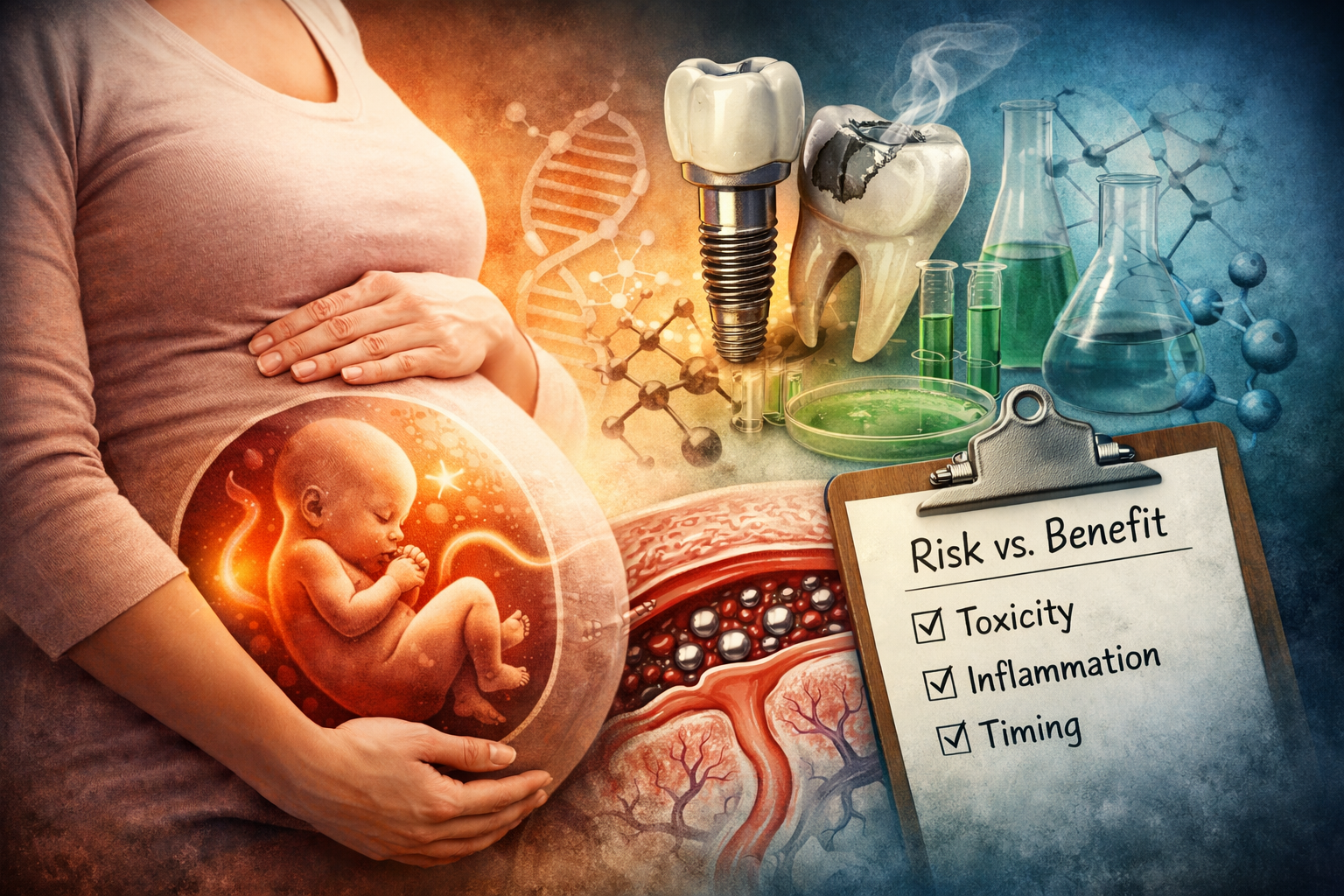
Pregnancy and Dental Toxicology: What Must Be Minimized, What Must Be Managed, and Why Timing Matters
Biological dentistry exists because a small number of clinicians were willing to confront a truth that conventional dentistry avoided for decades: dental materials are not biologically inert.
That truth, however, is only half the story. Biological dentistry also requires restraint — an understanding that there are moments when minimizing intervention is the most biologically responsible choice. Nowhere is that more important than during pregnancy and breastfeeding, when the developing nervous system of another human being is directly affected by maternal exposures. This article exists to clarify what matters most during pregnancy when it comes to dental toxicology — not to promote fear, purity, or unrealistic ideals, but to support informed, biologically grounded decision-making.
By that same token, I'm going to say something that I've said before. I'm a big believer in making standards that protect the most vulnerable members of our population. I think if we create standards to protect the "canaries" in societal "coal mines", EVERYONE will benefit. The unborn and newly born are society's "canaries". Our standards should reflect THEIR vulnerabilities and sensitivities. They do not. Hence the purpose of this article. Because if society won't protect them, then we must as individuals and biological dentists.
A Necessary Starting Point: Dentistry Is Prosthetic by Nature
There is no such thing as “organic” dentistry. Dentistry exists because biology sometimes fails:
teeth decay
enamel fractures
jaws don’t always grow correctly
tissues age
trauma happens
Every restoration is a compromise. Every material is man-made. Even the materials God made — enamel and dentin — do not last forever. So the goal of biological dentistry cannot be elimination of materials. It must be harm reduction, material selection, dose awareness, and timing.
Pregnancy is one of the clearest examples of why timing matters.
Pregnancy Changes the Rules
Pregnancy is not a detoxification window. It is a containment window. During pregnancy:
the placenta does not block toxins — it actively transports many of them
fetal detoxification systems are immature
neurodevelopment is exquisitely sensitive to chemical disruption
small exposures can have disproportionate effects
This does not mean dentistry should be completely avoided during pregnancy. In fact, it's vital to practice rigorous prevention because that is also important for the developing baby. It means toxicology must be taken seriously, and yes, there are SOME things that MUST be completely avoided during pregnancy. We will talk about those things right here.
The Core Biological Principle: Absorbed Means Systemic — Always
A foundational misunderstanding in dentistry is the idea that some materials or agents are “topical” and therefore biologically insignificant. This is not how human physiology works - at least not in the mouth. If a substance:
is absorbed through oral mucosa
is swallowed in saliva
enters circulation
crosses the placenta
appears in breast milk
then it is systemic. Pregnancy exposes this reality immediately, because anything absorbed by the mother is delivered to the fetus — often at higher relative concentrations. Regulatory language does not change biology.
Mercury: The Boundary Case That Defines Pregnancy Restraint
Mercury remains the clearest, most extensively studied example of why pregnancy demands clinical restraint in dentistry. Not because mercury is the only toxic material used in dental care — but because its toxicokinetics during pregnancy are so well understood that they remove any ambiguity about risk.
Mercury exposure increases during amalgam removal
Mercury crosses the placenta efficiently
Fetal mercury levels often exceed maternal levels
Mercury accumulates in fetal brain tissue
Even transient maternal spikes translate into fetal exposure
This is why mercury amalgam removal must not be performed during pregnancy or breastfeeding, regardless of protocol. SMART/PROTECT reduces exposure — it does not eliminate it. Pregnancy is not the time to mobilize mercury. This principle becomes the reference point for evaluating other materials.
The Mercury Science
Dental amalgam is approximately 50% elemental mercury by weight. When amalgams are drilled or removed, mercury vapor is released and rapidly absorbed through the lungs into systemic circulation. Multiple human studies have documented that mercury exposure increases acutely during amalgam removal, even when standard precautions are used. Here are some scientific studies to show this evidence in abundance. I want there to be NO question in anyone's mind once they read this information, because the vast majority of dentists today are still drilling amalgam on pregnant women without even thinking that they are in any way causing harm.
1. Sandborgh-Englund et al. (PMID: 9539465)
https://pubmed.ncbi.nlm.nih.gov/9539465
Removing all amalgam fillings at once caused a 32% increase in mercury levels in blood and plasma within 48 hours, but levels went down over the next few months. This means removal causes a temporary spike in mercury in the body.
2. Clarkson et al. (PMID: 14585942)
https://pubmed.ncbi.nlm.nih.gov/14585942/
Dental amalgam fillings release mercury vapor into the mouth and are the main source of mercury exposure for most people. Having 10 amalgam surfaces roughly doubles mercury levels in urine compared to people without fillings.
3. Jirau-Colón et al. (PMID: 30909378)
https://pubmed.ncbi.nlm.nih.gov/30909378
Mercury is highly toxic, and dental amalgam fillings constantly release mercury vapor that people breathe in. The review discusses concerns about mercury from fillings potentially linking to diseases like Alzheimer's and Parkinson's.
4. Björkman et al. (PMID: 36383213)
https://pubmed.ncbi.nlm.nih.gov/36383213
After removing all amalgam fillings, mercury and silver levels in blood dropped significantly within one year. Patients also reported feeling better, though it's unclear if mercury reduction directly caused the improvement.
5. Halbach et al. (PMID: 11032131)
https://pubmed.ncbi.nlm.nih.gov/11032131
Mercury levels in blood plasma peaked one day after amalgam removal, then decreased with half-lives of 3 and 43 days. Using a rubber dam during removal helped reduce mercury exposure during the procedure.
6. Ekstrand et al. (PMID: 9584901)
https://pubmed.ncbi.nlm.nih.gov/9584901
After removing all amalgam fillings, mercury levels in blood plasma dropped to 40% of original levels after 60 days. Mercury in feces increased dramatically for several days after removal, then gradually decreased.
7. Zwicker et al. (PMID: 25404430)
https://pubmed.ncbi.nlm.nih.gov/25404430
People with amalgam fillings had double the urine mercury compared to those without fillings. Removing fillings decreased mercury to normal levels and improved self-reported health symptoms compared to keeping the fillings.
8. Halbach et al. (PMID: 17767927)
https://pubmed.ncbi.nlm.nih.gov/17767927
After amalgam removal, inorganic mercury in blood dropped rapidly, stabilizing at 27% of pre-removal levels after 60 days. The estimated daily mercury dose absorbed from fillings was below safety limits set by health organizations.
9. Berglund & Molin (PMID: 9823089)
https://pubmed.ncbi.nlm.nih.gov/9823089
Using a rubber dam during amalgam removal significantly reduced the mercury spike in blood plasma. One year after removal, mercury levels in plasma were 52% lower and in urine were 76% lower than before removal.
10. Halbach et al. (PMID: 9600804)
https://pubmed.ncbi.nlm.nih.gov/9600804
Mercury in blood plasma peaked at 0.6 ng/ml after amalgam removal and decreased with half-lives between 5 and 13 days. Using a rubber dam during removal reduced the temporary mercury increase.
11. Geier (PMID: 41109985)
https://pubmed.ncbi.nlm.nih.gov/41109985
American adults with amalgam fillings had significantly higher blood mercury levels than those without fillings. More amalgam surfaces correlated with higher inorganic mercury in blood, showing a dose-dependent relationship.
12. Sinha et al. (PMID: 38699922)
https://pubmed.ncbi.nlm.nih.gov/38699922
Patients who had all amalgam fillings removed reported reduced intensity of both mouth-related and general health complaints at 1 and 5 years after removal compared to before removal.
Taken together, these studies demonstrate a consistent pattern:
mercury exposure increases during removal
the increase is usually transient
protective protocols reduce but do not eliminate exposure
For non-pregnant adults, this risk may be acceptable and manageable.
For pregnancy, it is not.
The Placenta Does Not Protect Against Mercury — It Actively Transfers It
One of the most critical — and least understood — facts about mercury toxicology is that the placenta is not a barrier. It is an exchange interface. Mercury crosses the placenta efficiently because it exploits amino acid transport systems intended to nourish the fetus.
1. Straka et al. (PMID: 26740192)
https://pubmed.ncbi.nlm.nih.gov/26740192
The placenta uses special amino acid transporters on its outer surface to actively bring methylmercury inside, then uses other transporters on the inner surface to push mercury toward the baby's side. This explains why mercury crosses the placenta so efficiently compared to other toxic metals.
2. Balthasar et al. (PMID: 28786956)
https://pubmed.ncbi.nlm.nih.gov/28786956
Methylmercury binds to cysteine (an amino acid) and looks similar to methionine (another amino acid). The placenta's amino acid transporters mistake mercury-cysteine for methionine and actively transport it across, which explains why mercury reaches the baby more efficiently than other heavy metals like lead or cadmium. This mechanism explains why fetal exposure is not merely proportional to maternal exposure — it is often amplified.
Fetal Mercury Levels Frequently Exceed Maternal Levels
Human biomonitoring studies consistently show that cord blood mercury levels equal or exceed maternal blood levels.
1. Sakamoto et al. (PMID: 29150340)
https://pubmed.ncbi.nlm.nih.gov/29150340
Mercury levels in cord blood (7.26 ng/g) were 1.9 times higher than in maternal blood (3.79 ng/g) in 54 Japanese mother-newborn pairs. Cord blood also had lower protective nutrients (vitamin E and DHA), suggesting fetuses face higher mercury toxicity risk than mothers.
2. Ou et al. (PMID: 24792881)
https://pubmed.ncbi.nlm.nih.gov/24792881
This meta-analysis found that fetuses are exposed to approximately twice as much methylmercury as mothers. The average cord-to-maternal blood ratio for methylmercury was 1.89, while inorganic mercury showed equal levels (ratio 1.01) between mother and baby.
3. Sakamoto et al. (PMID: 33640495)
https://pubmed.ncbi.nlm.nih.gov/33640495
In 55 Japanese mother-newborn pairs, mercury in cord blood red blood cells was 1.58 times higher than maternal levels (13.9 vs 9.16 ng/mL). Cord blood was rich in methylmercury, which easily transfers to the fetal brain, increasing toxicity risk.
4. Zhang et al. (PMID: 37625333)
https://pubmed.ncbi.nlm.nih.gov/37625333
The placental transfer ratio for mercury was 1.68, the highest among all toxic metals studied in 100 mother-newborn pairs. Mercury concentrations in late pregnancy strongly correlated with cord blood levels (correlation coefficient 0.66), showing efficient placental passage.
5. Kabamba & Tuakuila (PMID: 32569801)
https://pubmed.ncbi.nlm.nih.gov/32569801
This systematic review of 35 large studies found cord blood mercury levels were typically 1.34 times higher than maternal levels. Infants had 50-63% of the total mercury burden in the mother-baby pair, despite being much smaller.
6. Bocca et al. (PMID: 31374401)
https://pubmed.ncbi.nlm.nih.gov/31374401
Mercury and manganese in cord blood were about 2.0 times higher than maternal blood in 31 Italian mother-newborn pairs. Mercury showed strong correlation between maternal and cord blood, indicating fetal body burden reflects maternal exposure.
7. Dahiri et al. (PMID: 36099946)
https://pubmed.ncbi.nlm.nih.gov/36099946
Among 100 Spanish mother-newborn pairs, mercury showed the strongest correlation between maternal and cord blood (r = 0.779). This indicates efficient placental transfer, with fetal mercury levels closely tracking maternal exposure.
8. Kim et al. (PMID: 26516876)
https://pubmed.ncbi.nlm.nih.gov/26516876
In 104 South Korean pairs, cord blood mercury averaged 1.76 times higher than maternal blood, and methylmercury was 1.81 times higher. Unlike cadmium (which the placenta blocked), mercury accumulated in fetuses beyond maternal levels.
9. Obi et al. (PMID: 25112573)
https://pubmed.ncbi.nlm.nih.gov/25112573
Nigerian cord blood mercury averaged 1.5 times higher than maternal blood (5.1 vs 3.6 μg/L) in 95 pairs. Over one-third of participants exceeded EPA safety guidelines, showing significant prenatal mercury exposure in this population.
10. Kozikowska et al. (PMID: 23938449)
https://pubmed.ncbi.nlm.nih.gov/23938449
In 40 Polish women, 75% of cord blood samples exceeded the EPA safe dose (5.8 μg/L), with some reaching 17 μg/L. Positive correlations existed between mercury in placenta, umbilical cord, and cord blood.
11. Hadavifar et al. (PMID: 31813691)
https://pubmed.ncbi.nlm.nih.gov/31813691
Among 89 Iranian mother-infant pairs, there was a strong correlation (r = 0.890) between maternal and infant blood mercury. Mercury was measured in maternal blood, placental tissue, umbilical cord tissue, and cord blood across 360 samples.
These findings make one point unavoidable: Even modest maternal mercury exposure can result in disproportionately high fetal exposure.
Why This Matters: The Fetal Brain Cannot Detoxify Mercury
Once mercury crosses the placenta:
it accumulates in fetal brain tissue
the fetal nervous system lacks mature detoxification pathways
the blood–brain barrier is incompletely developed
As a result, exposure levels tolerated by adults can cause irreversible neurodevelopmental harm in the fetus. Large, well-designed human studies have repeatedly demonstrated associations between prenatal mercury exposure and:
reduced IQ
impaired attention and memory
deficits in language and visual-spatial processing
impaired motor development
1. Saavedra et al. (PMID: 33954792)
https://pubmed.ncbi.nlm.nih.gov/33954792
Summary: Prenatal mercury exposure was consistently linked to lower birth weight and worse scores on brain development tests in children up to age 8. Some protective nutrients in the mother's diet may reduce mercury's harmful effects.
2. Kim et al. (PMID: 29751424)
https://pubmed.ncbi.nlm.nih.gov/29751424
Summary: Korean babies whose mothers had higher mercury during early pregnancy scored lower on mental and motor development tests at 6 months old, even after accounting for beneficial nutrients from fish.
3. Dack et al. (PMID: 35206164)
https://pubmed.ncbi.nlm.nih.gov/35206164
Summary: This review of 32 studies found weak evidence linking prenatal mercury to poor brain development in children under 5. Most high-quality studies showed no clear connection between mercury and developmental delays.
4. Pan et al. (PMID: 34989572)
https://pubmed.ncbi.nlm.nih.gov/34989572
Summary: Mercury exposure during pregnancy causes long-term brain damage through oxidative stress, calcium imbalance, mitochondrial damage, and DNA changes. Babies and young children are much more sensitive to mercury than adults.
5. da Silva et al. (PMID: 40869893)
https://pubmed.ncbi.nlm.nih.gov/40869893
Summary: Review of 68 studies with 215,195 children found mercury exposure during pregnancy—especially early pregnancy—consistently harmed thinking, movement, and behavior. The developing brain is uniquely vulnerable even at low mercury levels.
6. Bjørklund et al. (PMID: 36063174)
https://pubmed.ncbi.nlm.nih.gov/36063174
Summary: Fetal brain damage can occur when maternal blood mercury is as low as 5 μg/L—a level mothers tolerate well. Mercury crosses the placenta and damages developing brain cells, causing lasting problems with movement and thinking.
7. Wang et al. (PMID: 30776746)
https://pubmed.ncbi.nlm.nih.gov/30776746
Summary: Low-level prenatal mercury exposure (average 2.00 μg/L in cord blood) was linked to poorer newborn brain function. The harmful effects were strongest in babies with lower DHA levels from their mothers.
8. Garí et al. (PMID: 34520749)
https://pubmed.ncbi.nlm.nih.gov/34520749
Summary: Polish school-age children exposed to higher mercury before birth had more hyperactivity and attention problems. Lead exposure was linked to lower IQ and worse math skills, while selenium and zinc appeared protective.
9. Ryu et al. (PMID: 28667852)
https://pubmed.ncbi.nlm.nih.gov/28667852
Summary: Children with higher mercury exposure during late pregnancy and early childhood showed more autistic behaviors at age 5. Doubling mercury levels increased autism-related social difficulties by 28-31%.
10. Kim et al. (PMID: 32871452)
https://pubmed.ncbi.nlm.nih.gov/32871452
Summary: Prenatal mercury exposure combined with rapid weight gain in the first 3 years caused worse brain development scores. This effect was strongest in children whose mothers had low folate during pregnancy.
11. Axelrad et al. (PMID: 17450232)
https://pubmed.ncbi.nlm.nih.gov/17450232
Summary: Combining data from three major studies, researchers found each 1 ppm increase in maternal hair mercury caused a 0.18 IQ point decrease in children. This relationship held across different populations and exposure levels.
12. O'Connor et al. (PMID: 39986421)
https://pubmed.ncbi.nlm.nih.gov/39986421
Summary: Review of 29 systematic reviews found prenatal mercury may increase risk of birth defects, preterm birth, ADHD, autism, and low birth weight. During childhood, higher mercury linked to ADHD and poor brain development.
All of this is documented developmental neurotoxicity. And yet, the US will not require dentists to stop using mercury amalgam in any way until 2034. Here is the article with that information. https://healthpolicy-watch.news/dental-amalgam-set-to-be-phased-out-by-2034-to-reduce-toxic-mercury-exposures/
YIKES. So the VAST majority of dentists are still blithely placing and removing amalgam on pregnant women and young children with zero regard for the studies that show the damage. Worse, there are rules that state that mercury amalgam should not be placed on pregnant women or children under age 6, and yet, I know for a FACT that amalgams are placed on young children and pregnant women all the time. I've seen it in some of the patients I have treated myself. But here is the link so you can see this for yourselves: https://www.healthaffairs.org/content/forefront/children-color-and-low-income-kids-still-receive-unsafe-mercury-based-dental-fillings
I'm also going to ask - What is the magic that happens between the last day a child is 5 years old, and the first day a child is 6 years old that makes it all of the sudden okay to place amalgam on the 6 year old? Because the rules the FDA has tells dentists to not use amalgam on children under age 6. Here is the link so that you can see for yourselves: https://www.fda.gov/medical-devices/dental-amalgam-fillings/information-patients-about-dental-amalgam-fillings#:~:text=Placement%20of%20new%20amalgam%20fillings,levels%20in%20umbilical%20cord%20blood.
Why Pregnancy Requires a Firm Boundary
Given what we know:
mercury exposure increases during amalgam removal
the placenta actively transfers and concentrates mercury
fetal mercury levels often exceed maternal levels
the developing brain cannot detoxify mercury
there is no biologically defensible argument for mobilizing mercury during pregnancy or breastfeeding. SMART and PROTECT protocols were developed to reduce exposure in non-pregnant adults. They significantly lower risk — but they do not eliminate it. No protocol can override placental transport biology. Even brief maternal mercury spikes translate into direct fetal exposure during critical windows of brain development. For this reason, mercury amalgam removal must not be performed during pregnancy or lactation — regardless of technique, intention, or protocol.
Pregnancy is not the time to place OR remove mercury from the body. It is the time to protect developing life. This boundary is not ideological. It is dictated by toxicology and placental physiology. And it is precisely this boundary that becomes the reference point for evaluating all other dental materials during pregnancy.
Fluoride: The Myth of “Topical” Exposure in Pregnancy
If mercury defines the absolute boundary in pregnancy, fluoride defines the most persistent misunderstanding. Fluoride is often described as “topical” in dentistry — a word that implies:
local effect
minimal systemic exposure
negligible risk beyond the tooth surface
That framing is biologically false. Fluoride is not topical in any meaningful toxicologic sense.
It is readily absorbed through oral mucosa, gastrointestinal tract, skin, and lungs — and once absorbed, it is systemic. There is no such thing as “topical-only fluoride exposure.”
1. Lockner F, et al. PMID: 28105705
https://pubmed.ncbi.nlm.nih.gov/28105705
Scientists measured fluoride in children's urine after applying fluoride varnish to their teeth. Urine fluoride levels significantly increased after varnish application, showing the body absorbed and then eliminated fluoride through urine. Some children had higher-than-ideal fluoride exposure.
2. Twetman S, et al. PMID: 10614903
https://pubmed.ncbi.nlm.nih.gov/10614903
Researchers measured fluoride in saliva from different glands after applying fluoride varnish. Fluoride levels increased in saliva for up to 6 hours, showing fluoride was absorbed and secreted back into the mouth through saliva glands.
3. Lin YS, et al. PMID: 30803490
https://pubmed.ncbi.nlm.nih.gov/30803490
Scientists measured silver and fluoride in blood after applying silver diamine fluoride to adults' teeth. Blood fluoride fluctuated around normal levels, and fluoride appeared in urine, confirming some absorption occurred but remained safe.
4. Ellenikiotis H, et al. PMID: 35484770
https://pubmed.ncbi.nlm.nih.gov/35484770
Researchers measured silver and fluoride in children's blood after applying silver diamine fluoride to cavities. They detected measurable silver in blood but no clear pattern for fluoride, confirming systemic absorption occurs safely.
5. Zero DT, et al. PMID: 1401438
https://pubmed.ncbi.nlm.nih.gov/1401438
Scientists measured fluoride in saliva from salivary glands after people used fluoride toothpaste, rinse, or gel. Fluoride levels increased slightly in gland saliva, showing fluoride was absorbed and secreted back through glands.
Why This Matters in Pregnancy
Pregnancy changes fluoride toxicokinetics in ways that increase fetal exposure:
Fluoride crosses the placenta
Fetal fluoride levels reflect maternal exposure
The developing brain is uniquely vulnerable
There is no detoxification advantage for the fetus
Effects occur at exposure levels previously considered “safe”
This places fluoride squarely in the category of developmental neurotoxicants — not because it causes acute poisoning, but because it subtly interferes with brain development during critical windows. And subtle neurotoxicity is precisely what pregnancy toxicology exists to prevent.
Fluoride Crosses the Placenta — Easily and Consistently
Human studies have repeatedly demonstrated that fluoride passes freely from mother to fetus.
Fluoride is not filtered out.
It is not buffered.
It is not “blocked.”
Maternal plasma fluoride levels correlate directly with:
placental fluoride concentration
cord blood fluoride
fetal tissue exposure
Representative Studies
1. Bashash et al. (2017) . PMID: 28937959
https://pubmed.ncbi.nlm.nih.gov/28937959
This study from Mexico found that when pregnant mothers had higher fluoride levels in their urine, their children scored lower on intelligence tests at ages 4 and 6-12. For every 0.5 mg/L increase in maternal urinary fluoride, children's IQ scores dropped by about 2.5-3 points.[3]
2. Green et al. (2019) . PMID: 31424532
https://pubmed.ncbi.nlm.nih.gov/31424532
This Canadian study measured fluoride in pregnant women's urine and found that boys (but not girls) had lower IQ scores when their mothers had higher fluoride exposure. A 1 mg/L increase in maternal urinary fluoride was linked to a 4.5-point decrease in boys' IQ scores at ages 3-4.[4]
3. Malin & Till (2015)
Exposure to fluoridated water and ADHD prevalence . PMID: 26001814
https://pubmed.ncbi.nlm.nih.gov/26001814
This population-based study found higher rates of ADHD in areas with fluoridated water, suggesting early-life fluoride exposure may impact neurodevelopment.
4. Goodman et al. (2022) - ELEMENT Cohort Longitudinal Analysis . PMID: 35276192
https://pubmed.ncbi.nlm.nih.gov/35276192
This study followed children from the same Mexican cohort as Bashash et al. across three time points (ages 4, 5, and 6-12 years). Each 0.5 mg/L increase in maternal urinary fluoride was associated with a 2.12-point decrease in overall IQ and a 2.63-point decrease in Performance IQ (visual-spatial and perceptual reasoning abilities). The negative association remained consistent across childhood, suggesting prenatal fluoride exposure has persistent effects on non-verbal intelligence.
5. Farmus et al. (2021) - Critical Windows Study . PMID: 34051202
https://pubmed.ncbi.nlm.nih.gov/34051202
This Canadian study examined 596 mother-child pairs to identify when children are most vulnerable to fluoride neurotoxicity. The prenatal window showed the strongest association with Performance IQ deficits (B = -2.36 per standardized unit), particularly in boys, though infancy was also a critical period. The study demonstrated that timing of exposure matters, with prenatal exposure showing the most pronounced effects.
6. Malin et al. (2024) - MADRES Cohort . PMID: 38767917
https://pubmed.ncbi.nlm.nih.gov/38767917/
This is the first U.S.-based prospective cohort study examining prenatal fluoride and neurobehavior, following 229 mother-child pairs in Los Angeles. Each 0.68 mg/L increase in maternal urinary fluoride was associated with nearly double the odds of neurobehavioral problems being in the borderline clinical or clinical range at age 3 years, plus a 2.29-point increase in internalizing problems (anxiety, emotional reactivity, somatic complaints).
7. Dewey et al. (2023) - Calgary Natural Experiment . PMID: 37236475
https://pubmed.ncbi.nlm.nih.gov/37236475
This unique study leveraged Calgary's 2011 decision to stop water fluoridation, comparing 616 children whose mothers were fully exposed, partially exposed, or not exposed to fluoridated water (0.7 mg/L) during pregnancy. Full prenatal exposure was associated with poorer executive function, particularly inhibitory control and cognitive flexibility in girls at ages 3-5 years, even though no differences in overall IQ were found.
8. Till C, Green R, Flora D, et al. PMID: 31743803
https://pubmed.ncbi.nlm.nih.gov/31743803
This Canadian study found that babies fed formula mixed with fluoridated tap water had lower IQ scores at ages 3-4 compared to breastfed babies. For every 0.5 mg/L increase in water fluoride (the difference between fluoridated and non-fluoridated areas), formula-fed children scored 9 points lower on performance IQ tests.
9. Taylor et al. (2025) - JAMA Pediatrics Systematic Review. PMID: 39761023
https://pubmed.ncbi.nlm.nih.gov/39761023/
This comprehensive meta-analysis of 74 studies found an IQ decrease of 1.63 points per 1 mg/L increase in urinary fluoride across all studies, with a 1.14-point decrease when restricted to low risk-of-bias studies. The inverse association persisted even when exposure was restricted to lower levels, including below 1.5 mg/L in some analyses.
These studies collectively demonstrate that prenatal fluoride exposure at levels common in fluoridated communities is associated with measurable neurodevelopmental impacts, including lower IQ scores, executive dysfunction, and increased neurobehavioral problems. Fluoride
reaches the fetus
accumulates during development
is associated with measurable neurodevelopmental harm
“Topical” Is a Marketing Term — Not a Biological One
Dentistry often reassures patients that fluoride varnish, gel, or toothpaste is “topical” and therefore safe. That reassurance collapses under even minimal scrutiny. Fluoride:
penetrates oral mucosa
is swallowed in measurable amounts
is absorbed systemically
enters circulation within minutes
As seen above in the multiple studies, they found that measuring plasma and urinary fluoride after “topical” application shows systemic uptake every time. Calling fluoride “topical” is like calling nicotine gum “topical.” It may be applied locally, but its effects are systemic.
The Developing Brain Is Not a Small Adult Brain
Fluoride neurotoxicity does not require high-dose exposure.
The fetal brain:
has incomplete blood–brain barrier function
is undergoing rapid neuronal migration and synapse formation
is exquisitely sensitive to endocrine and oxidative disruption
Fluoride has been shown to:
interfere with thyroid hormone signaling
induce oxidative stress
alter neurotransmitter levels
disrupt hippocampal development
These are not theoretical risks. They are documented mechanisms.
Regulatory “Safety” Is Not Pregnancy-Specific Safety
One of the most misleading aspects of fluoride policy is that safety thresholds are based on:
adult toxicity
skeletal fluorosis
dental fluorosis
They are not based on fetal neurodevelopment. Much like mercury, fluoride safety limits were set before modern developmental neurotoxicity testing existed — and they have not meaningfully incorporated newer evidence. So when someone says: “Fluoride is safe at regulated levels”
The appropriate follow-up question is: “Safe for whom — and at what stage of development?”
Because pregnancy, breastfeeding infants and children are not included in that reassurance.
Fluoride During Pregnancy: The Biological Conclusion
Given what we know:
Fluoride is systemically absorbed
Fluoride crosses the placenta
Fetal exposure reflects maternal exposure
Neurodevelopmental effects occur at common exposure levels
There is no fetal detoxification advantage
There is no biologically defensible reason to have fluoride exposure during pregnancy. In all honesty, I don't believe fluoride exposure is necessary at ANY phase of life, except perhaps in the fluoride gases for general surgery, and that is only until they come up with something better. Truly.
Just as pregnancy is not the time to mobilize mercury, it is not the time to add unnecessary systemic toxicants. But particularly under the guise of prevention, which is how it is sold to the entire population of this planet.
Where This Fits in Biological Dentistry
Biological dentistry does not promise “zero exposure.” That would be dishonest. What it does promise is risk prioritization. During pregnancy, the priority is clear:
protect neurologic development
minimize avoidable systemic toxicant exposure
avoid interventions that offer marginal benefit with disproportionate fetal risk
Fluoride fails that test during pregnancy. And once we understand that fluoride is never truly topical, and it can cause harm to developing brains, we must ask ourselves why we would want to ever expose ourselves to fluoride at ANY age. Because the question again becomes - at what magical age does fluoride become safe for the brain? What is the difference between age 5 and 364 days and age 6 and 1 day?
BPA & Resin-Based Dental Materials: Endocrine Disruption in Disguise
If mercury is the boundary case and fluoride is the marketing myth, BPA is the Trojan horse of modern dentistry. Bisphenol A (BPA) is not a fringe contaminant. It is a known endocrine-disrupting chemical used in the manufacture of many resin-based dental materials — including:
composite restorations
bonding agents
sealants
orthodontic adhesives
some provisional materials
And unlike mercury, BPA does not announce itself as dangerous. It is colorless. Odorless. Esthetic. Marketed as “modern.” Which is precisely the problem. Modern dentistry relies heavily on resin-based materials. These materials have real advantages — but they are not biologically inert. MOST dental resins and bonding agents:
release bisphenol analogs (including BPA derivatives)
act as endocrine disruptors
interfere with estrogen signaling
are absorbed systemically, particularly when uncured monomers are present
BPA Is Not Inert — It Is Biologically Active at Extremely Low Doses
BPA is a xenoestrogen — a synthetic compound that mimics estrogen and interferes with hormonal signaling. This matters profoundly in pregnancy, because fetal development is orchestrated by exquisitely timed hormonal cues. You do not need high doses to disrupt that choreography. In fact, low-dose exposure is often more disruptive than high-dose exposure due to non-linear endocrine effects. This is not controversial toxicology. This is settled science.
Dental Materials Are a Documented Source of BPA Exposure
Many dental composites and sealants do not contain BPA itself — but they contain BPA derivatives such as:
Bis-GMA
Bis-DMA
Bis-EMA
These compounds degrade into BPA through:
enzymatic activity in saliva
hydrolysis
mechanical wear
incomplete polymerization
Which means BPA exposure is not hypothetical. It is measurable.
Representative Studies for BPA
1. Joskow et al. (2006) - BPA Exposure from Dental Sealants. PMID: 16570469
https://pubmed.ncbi.nlm.nih.gov/16570469
This study tested 14 men after getting dental sealants. One type of sealant (Delton LC) released measurable BPA into saliva and urine, with patients exposed to about 110 micrograms of BPA. Another sealant (Helioseal F) released almost no BPA. The study showed some dental sealants can be a source of BPA exposure.
2. Kingman et al. (2012) - BPA in Saliva and Urine After Composite Placement. PMID: 23204083
https://pubmed.ncbi.nlm.nih.gov/23204083
This study measured BPA in 172 people before and after getting composite fillings. BPA levels in saliva increased within one hour after the filling was placed but returned to normal within 8 hours. Urine BPA levels showed a 43% increase 9-30 hours after treatment.
3. Fleisch et al. (2010) - BPA and Related Compounds in Dental Materials. PMID: 20819896
https://pubmed.ncbi.nlm.nih.gov/20819896
This review found that BPA is released from dental sealants and fillings through breakdown of BPA-containing chemicals in saliva. BPA can be detected in saliva for up to 3 hours after placement. The authors recommend using these materials carefully, especially during pregnancy.
4. McKinney et al. (2020) - Prospective Study of BPA from Dental Treatment. PMID: 32579872
https://pubmed.ncbi.nlm.nih.gov/32579872
This study followed 211 children ages 4-12 who received composite fillings or sealants. Urinary BPA levels were 86% higher two days after dental treatment compared to before treatment. BPA levels returned to normal within 4 weeks. Children who had more tooth surfaces treated had higher BPA exposure.
The pattern is consistent:
BPA exposure spikes after placement
exposure is systemic
degradation continues over time
BPA Crosses the Placenta — and Alters Fetal Programming
Like mercury and fluoride, BPA does not stop at the placenta.
It crosses readily and accumulates in fetal tissues, where it interferes with:
estrogen signaling
thyroid hormone regulation
neurodevelopmental pathways
metabolic programming
Based on the medical literature, BPA crosses the placenta, accumulates in fetal tissues, and disrupts multiple hormonal and developmental pathways critical for fetal development. Here are studies demonstrating these mechanisms
Placental Transfer and Fetal Accumulation:
1. Nesan et al. (2018) - BPA Crosses the Placental Barrier. PMID: 29241697
https://pubmed.ncbi.nlm.nih.gov/29241697
BPA crosses the human placental barrier and has been found in the blood of human fetuses during pregnancy. The brain is a key target for BPA disruption during development, leading to behavioral disorders later in life.
2. Vom Saal (2016) - BPA Fetal Programming Effects. PMID: 27482660
https://pubmed.ncbi.nlm.nih.gov/27482660
Very low doses of BPA during fetal life cause permanent changes in organ development and function. Small shifts in estrogen and testosterone levels from BPA exposure can affect organs throughout life and increase disease risk in adulthood.
Estrogen Signaling Disruption:
3. Vom Saal & Vandenberg (2021) - BPA Endocrine Disruption Mechanisms. PMID: 33516155
https://pubmed.ncbi.nlm.nih.gov/33516155
BPA acts like estrogen in the body and causes harmful effects at extremely low doses (as low as 2.5 micrograms per kilogram per day). Traditional safety testing methods fail to detect BPA's endocrine-disrupting effects because they don't account for how hormones actually work.
4. Cimmino et al. (2020) - BPA Hormone Receptor Binding
PMID: 32796699
https://pubmed.ncbi.nlm.nih.gov/32796699
BPA binds to estrogen receptors and androgen receptors, mimicking hormones in the body. This affects body weight, cancer development, metabolism, reproductive function, and causes epigenetic changes like DNA methylation that can alter how genes are expressed.
Thyroid Hormone Disruption:
5. Ahmed (2016) - BPA Disrupts Fetal Thyroid Function. PMID: 27326465
https://pubmed.ncbi.nlm.nih.gov/27326465
When pregnant rats were exposed to BPA, both mothers and fetuses had lower thyroid hormone levels (T3 and T4) and higher TSH. Fetal thyroid glands showed tissue damage including swelling and abnormal cell growth. This disrupted growth hormones and metabolism in the developing fetuses.
Neurodevelopmental Pathway Disruption:
6. Henriksen et al. (2020) - BPA Alters Brain Gene Expression. PMID: 32365465
https://pubmed.ncbi.nlm.nih.gov/32365465
BPA exposure during pregnancy changed the expression of 259 genes in the developing brain's hypothalamus. Many of these genes are involved in autism spectrum disorders, brain cell development, hormone signaling, learning, and behavior. Fifteen genes linked to autism were specifically affected.
7. Welch & Mulligan (2022) - BPA Neurodevelopmental Toxicity. PMID: 35270035
https://pubmed.ncbi.nlm.nih.gov/35270035
Studies in animals show BPA exposure during development impairs brain stem cell growth, synapse formation (connections between brain cells), and learning. BPA-exposed animals show altered movement, memory problems, and anxiety-like behavior—similar to symptoms seen in ADHD and autism.
Account required
Sign in to see more
Create an account or sign in to unlock the full list.