Resource Libraries
Types of Dental Bone Grafts: What Is Placed in Your Body?
Types of dental bone grafts, from autograft and allograft to bovine xenograft and synthetic options, why bone loss starts after extraction, and what to ask.
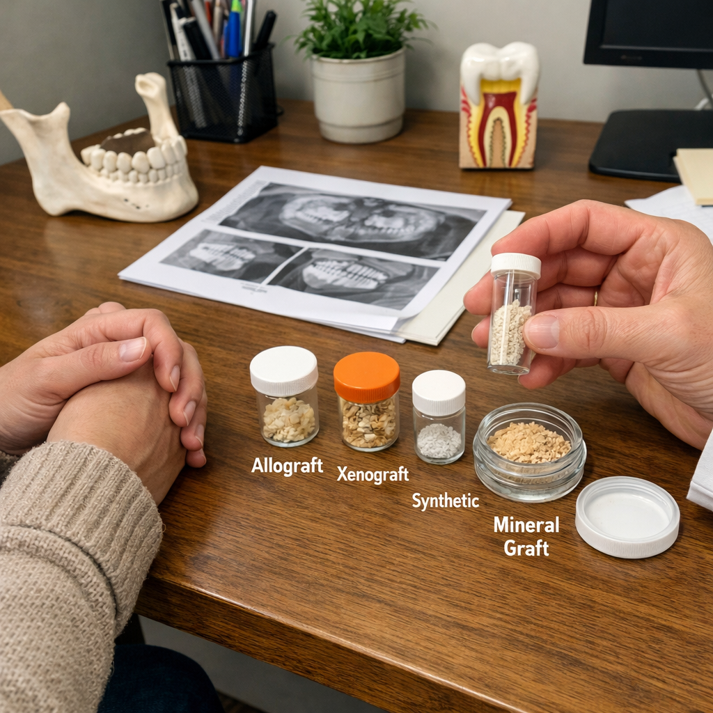
Bone Grafts in Dentistry: What They Are, Why They Matter, and How to Understand What Is Being Placed in Your Body
Bone grafting is not one of the most common procedures in dentistry — and that is exactly the problem. If you have read any of the articles on this platform discussing bone loss after extraction, jawbone cavitations, or the newer term Covered Socket Residuum (CSR), you already understand the core issue: how a socket heals matters. Bone loss after extraction is not rare, unexpected, or a complication. It is expected biology. Despite decades of research showing that bone loss begins immediately after a tooth is removed, many teeth are still extracted with no attempt to preserve the surrounding bone. This is not because bone grafting is unimportant. It is because it is often:
-
not covered by insurance
-
not emphasized as routine
-
not clearly explained to patients
-
dentist's lack of training on grafting as it's not taught in dental school
-
dentists confusion regarding types of grafting options
As a result, grafting is frequently framed as optional, elective, or cosmetic — when in reality it influences long-term jaw structure, implant stability, facial support, bite mechanics, and in some patients, airway integrity. Bone loss after extraction is not a failure of dentistry. It is the predictable biological response to loss of stimulation. Bone grafting, when done appropriately, is a way to work with biology instead of passively accepting preventable loss.
What This Article Is Designed to Do
This article has three goals:
-
Explain what bone grafts are and how they work
-
Explain why biocompatibility and compatibility testing matter
-
Clearly outline the categories of bone graft materials and the actual brands patients encounter in the United States, Europe, and Australia
-
I'm going to give you the science, but due to the sheer amount, I'm only listing the PMIDs. It's a departure from my usual style, but when I tried to do it the way I normally do it, I realized it was going to end up confusing my readers. If you really want the science, please by all means copy and paste the PMID into your search bar and google it. If I have a typo and the PMID doesn't match, please contact Kim at Admin@OraBiologics.com and she will let me know to correct it.
This is a HUGE and complex topic. The scientific literature is extensive. In order to remain accurate without oversimplifying, each graft category is presented using the same structure so that risks, benefits, pros, cons, and consequences can be clearly compared and contrasted.
What Is a Bone Graft?
Bone is living tissue. It is not inert. It constantly responds to:
-
mechanical load (chewing forces)
-
blood supply
-
inflammation
-
hormones
-
immune system signaling
When bone is lost due to extraction, infection, trauma, or chronic inflammation, the adult body often does not fully regenerate it on its own. A bone graft does not magically “turn into bone.”
Instead, a bone graft supports healing by doing one or more of the following:
-
Providing a structure that new bone can grow along
-
Providing biological signals that encourage bone formation
-
Providing living cells capable of forming bone
Different graft materials do different combinations of these things. That distinction is critical.
How Bone Grafts Work: Three Concepts That Explain Everything
These terms sound technical, but the concepts are simple.
-
Osteoconduction — “Structure”. The graft provides a scaffold. Bone can grow along it, but the graft itself does not tell the body what to do.
-
Osteoinduction — “Instructions”. The graft provides biological signals that encourage the body to make bone.
-
Osteogenesis — “Living Builders”. The graft contains living bone-forming cells.
Only autografts (material taken from your own body) reliably provide all three. Most dental bone grafts provide structure only and rely entirely on the patient’s biology to do the rest.
Redefining “Success” in Bone Grafting
Many grafts look similar on X-rays. Dense white areas are often described as “successful bone.”
That is an incomplete definition. A more biologically meaningful definition of success includes:
-
what the tissue looks like under a microscope
-
whether the graft remodels into native bone or remains as foreign material
-
whether the immune system tolerates it quietly or remains chronically activated
-
long-term function (implant stability, bite support, facial structure)
-
broader structural consequences, including airway support when relevant
When success is defined this way, it becomes obvious why no graft is universally best.
Why Biocompatibility and Compatibility Testing Matter
Biocompatibility is often treated as a yes-or-no concept. In reality, it exists on a spectrum.
Every material placed in the body interacts with the immune system. Some materials are quietly tolerated. Others provoke ongoing immune vigilance, even in the absence of infection.
This matters especially for people with:
-
autoimmune disease
-
chronic inflammation
-
diabetes
-
smoking history
-
airway compromise
-
oral microbiome imbalance
-
previous implant or graft failures
-
gut absorption issues
-
nutrtional status
-
vitamin D levels
-
hormone imbalance or deficiency
In many cases, patient biology matters more than the brand name. This is why compatibility testing matters. No material is universally compatible with every person. Clinical experience consistently shows that patients tend to do best with materials their body accepts — even when those materials are not theoretically “ideal” on paper. Biology does not respect dogma. Unfortunately, biocompatibility testing is not widely available for bone grafting materials. So some of this science is difficult for me to...process, because I think so much depends on compatibility, and it's just not done. I would bet those numbers change with compatibility testing. And as we have learned to understand with root canals, just because something is tolerated by the body doesn't really mean it's fully accepted. It may drive inflammation further on down the line. There is still so much to study.
A Perspective Many Holistic Consumers Share
COVID Vaccination, Donor Tissue, and Why Some People Choose to Avoid Allografts
For many patients in holistic and biologically oriented communities, the COVID era fundamentally changed how they think about medical risk, informed consent, and donor-derived materials. A significant number of people have personally witnessed serious illness, autoimmune conditions, cardiovascular events, aggressive cancers, or death following COVID vaccination in friends, family members, or patients. For those individuals, this concern is not theoretical — it is lived experience.
From that perspective, it is reasonable to ask: If disease transmission from human donor tissue is not zero risk, and if most donors are now vaccinated against COVID, is it appropriate for me to accept human donor bone graft material at all? That question deserves to be addressed honestly — not dismissed. Even outside of COVID-related concerns, human allografts have never been zero risk:
-
donor tissue comes from another person
-
screening and processing reduce risk dramatically
-
but they do not eliminate risk entirely
Another practical reality: most tissue banks do not provide donor COVID vaccination status as a selectable criterion for dental bone grafts. That means:
-
patients cannot reliably request “unvaccinated donor bone”
-
dentists often cannot verify donor vaccination status
-
choosing an allograft usually involves accepting uncertainty
For patients who are uncomfortable with that uncertainty, the ethical response is not reassurance — it is choice. At the same time, I also want to share something from my own clinical experience: in our office, when we perform compatibility testing, many patients test compatible with at least one of , but usually 2 or 3 of our allograft options. We also see compatibility with synthetic options — but less frequently in our patient population. That doesn’t mean one category is “good” and one is “bad.” It means individual biology matters, and it’s one more reason we don’t treat humans like cookie cutters.
Is There a “Best” Bone Graft?
There is no single graft that is best for every patient and every situation.
From a biological standpoint, however, autografts occupy a unique reference position. Understanding autografts first makes every other graft category easier to evaluate.
CATEGORY 1: AUTOGRAFTS - Bone Grafts From Your Own Body
A. What an Autograft Is
An autograft is bone graft material taken from the patient’s own body and placed elsewhere in that same patient. In dentistry, this can include:
-
bone harvested from another area of the jaw during surgery
-
tooth-derived graft material created from the patient’s own extracted tooth
B. What Autografts Do Biologically
Autografts are unique because they can provide:
-
Osteoconduction (structure)
-
Osteoinduction (biological signaling)
-
Osteogenesis (living bone-forming cells)
This combination makes autografts the biological reference standard against which all other grafts are compared.
Supporting science: PMIDs: 37297251, 35384291, 41607115, 27915981, 30633080, 33563416, 10693546
C. Clinical Strengths (Pros)
Autografts consistently demonstrate:
-
the highest percentage of new bone formation
-
predictable biological integration
-
absence of foreign-body immune response
They are considered the gold standard for bone regeneration because they actively participate in healing rather than merely supporting it.
Supporting science: PMIDs: 37297251, 27915981, 30633080
D. Limitations (Cons)
Autografts also have real drawbacks:
-
limited quantity available in the mouth
-
need for a second surgical site (when harvesting bone)
-
donor-site pain, swelling, or complications
-
increased surgical time
These limitations often make autografts impractical for routine socket grafting in everyday dentistry.
Supporting science: PMIDs: 30633080, 33563416, 10693546
E. Immune and Safety Considerations
Autografts have zero immunogenicity because the tissue originates from the patient’s own body. There is:
-
no risk of immune rejection
-
no disease transmission risk
-
no donor variability
This makes autografts biologically ideal — when they are feasible.
Supporting science: PMIDs: 10693546, 33563416
F. Forms and Subtypes Patients May Encounter
Autografts in dentistry have two meaningful subtypes:
-
Autogenous bone (harvested from the patient’s jaw during surgery)
-
Tooth-derived autograft (autogenous dentin graft from an extracted tooth)
Tooth-derived autografts use dentin, which shares similar mineral and collagen composition with bone, explaining their biological compatibility. Autogenous dentin grafts demonstrate:
-
osteoconductive and osteoinductive behavior
-
favorable histologic remodeling
-
ridge preservation comparable to conventional grafts
-
gradual resorption and replacement with native bone
Resorption timing varies (approximately 2–24 weeks), which can be an advantage or limitation depending on the clinical goal.
Supporting science: PMIDs: 38179035, 27279547, 32664303, 38050282, 41598544, 36574044, 29095791, 36739203
G. Common Clinical Uses (Indications)
Autografts are most often used when maximum regeneration is needed and the clinical team accepts higher complexity, including:
-
ridge augmentation (especially where rapid remodeling is desired)
-
localized defect repair during implant surgery
-
cases where immune reactivity must be minimized
-
tooth-derived grafting for socket preservation when an extraction is already planned
(Autografts can be used for routine socket grafting, but logistical limitations often prevent this in day-to-day dentistry.)
Supporting science: PMIDs: 37297251, 27915981, 38179035, 38050282
H. Cost and Practical Considerations
Autografts often increase:
-
surgical time
-
surgical complexity
-
patient morbidity (especially if a donor site is required)
Tooth-derived autografts can reduce donor-site morbidity because the tooth is already being removed, but they still require:
-
a suitable tooth
-
processing time and equipment
-
consistent protocol execution
Supporting science: PMIDs: 30633080, 33563416, 29095791
I. Brands / Systems in the US, Europe, Australia
For autogenous bone harvested from the patient, there are no commercial brands — this is technique-based. For tooth-derived autografts, chairside processing systems commonly include:
-
KometaBio (Smart Dentin Grinder)
-
Tooth Transformer
-
AutoBT
-
BonMaker
These systems differ in cleaning, grinding, and demineralization protocols. Outcomes depend more on technique and biology than branding.
J. Trade-Off Statement (Compare / Contrast)
Autografts provide the most complete biology (structure + signals + living cells) but are limited by availability and surgical burden.
-
outperform allografts biologically but require more invasiveness
-
remodel more completely than xenografts but resorb faster
-
are more biologically active than naturally derived minerals and synthetics, which function primarily as scaffolds
K. Science Supporting Autografts (Complete List)
Autogenous bone
-
Ferraz MP. PMID: 37297251
-
Zufía J, Abella Sans F. PMID: 35384291
-
Palombo D, et al. PMID: 41607115
-
Lu J, et al. PMID: 27915981
-
Baldwin P, et al. PMID: 30633080
-
Schmidt AH. PMID: 33563416
-
Bauer TW, Muschler GF. PMID: 10693546
Tooth-derived autografts (autogenous dentin)
-
Hussain AA, et al. PMID: 38179035
-
Pang KM, et al. PMID: 27279547
-
Sánchez-Labrador L, et al. PMID: 32664303
-
Feng Y, et al. PMID: 38050282
-
Jáquez M, et al. PMID: 41598544
-
Barreiro BOB, et al. PMID: 36574044
-
Gharpure AS, Bhatavadekar NB. PMID: 29095791
-
Abo-El-Saad MM, et al. PMID: 36739203
CATEGORY 2: ALLOGRAFTS - Human Donor Bone Grafts
A. What a Human Bone Allograft Is
A human bone allograft is bone that comes from another human being, not from the patient. The bone is donated, screened, processed by a tissue bank, and then used to support bone regeneration in dental procedures. Allografts are human-derived, but they are not living tissue by the time they are placed.
Supporting science: PMIDs: 24046051, 22632396
B. What Allografts Do Biologically
Allografts primarily work through osteoconduction, providing a three-dimensional scaffold for the patient’s bone to grow into. Depending on processing, some allografts may retain osteogenic signaling proteins (including BMPs), but the amount and activity are variable across products.
Allografts do not contain living bone-forming cells.
Supporting science: PMIDs: 38068918, 22632396, 35816626
C. Clinical Strengths (Pros)
Allografts are widely used because they:
-
avoid a second surgical site
-
are available in many forms for different clinical needs
-
can achieve comparable clinical outcomes to autografts in certain applications (especially socket preservation and moderate ridge augmentation)
Supporting science: PMIDs: 24046051, 22632396, 22559727, 22697457, 38068918
D. Limitations (Cons)
Allografts have limitations including:
-
lack of living cells (reduced osteogenesis compared to autograft)
-
variable performance depending on processing method
-
less predictable outcomes in certain demanding procedures (especially vertical augmentation with blocks)
-
remodeling behavior that can be inconsistent between products
Supporting science: PMIDs: 35816626, 22559727, 31828520
E. Immune and Safety Considerations
Allografts introduce foreign human tissue. Modern processing reduces antigenicity, but immune recognition is not always eliminated. Evidence shows:
-
measurable immune responses occur in a substantial subset of patients (often cited around 48%)
-
serious local complications remain rare
-
the long-term significance of persistent low-grade immune signaling is not fully known
Disease transmission risk is considered very low due to donor screening, serologic testing, and processing protocols — but it is not zero. Studies on commercial products have found reassuring immune marker findings in some cases (e.g., no detectable MHC class I residues and no major inflammatory marker differences).
Supporting science: PMIDs: 22697457, 24046051, 22559727, 31424173
F. Forms and Subtypes Patients May Encounter
Allografts are sold in meaningful subtypes that affect behavior:
-
Mineralized vs demineralized (demineralized forms may expose more signaling proteins, but results vary)
-
Cortical vs cancellous
-
Particulate vs block
These distinctions matter clinically: blocks behave differently than particulates, and processing influences mechanical integrity, remodeling speed, and biologic signaling.
Supporting science: PMIDs: 22559727, 37099570, 35816626, 38068918
G. Common Clinical Uses (Indications)
Allografts are commonly used for:
-
socket preservation
-
ridge augmentation (moderate defects)
-
sinus elevation (alone or mixed)
-
horizontal and vertical augmentation (often with barrier membranes)
Important limitation: allogeneic blocks show higher failure rates and more fibrous tissue than autogenous blocks in vertical augmentation.
Supporting science: PMIDs: 22632396, 31828520
H. Cost and Practical Considerations
Allografts often reduce:
-
operative time
-
surgical complexity
-
patient morbidity
However, material cost can be higher than some synthetic alternatives, and pricing varies by form and processing method.
Supporting science: PMID: 24046051
I. Brands / Systems in the US, Europe, Australia
Patients may encounter allografts from:
-
Maxxeus
-
Karl Schumacher
-
PuraGraft
-
LifeNet Health (OraGRAFT®)
-
Straumann (maxgraft®)
-
botiss biomaterials
-
ZimVie (Puros®)
-
Nobel Biocare (creos™ allo.gain)
-
BioHorizons
-
CAMLOG
These companies may sell multiple formulations, so the brand name alone does not tell you whether the graft is mineralized, demineralized, particulate, or block.
J. Trade-Off Statement (Compare / Contrast)
Allografts reduce surgical burden while providing solid osteoconductive scaffolding, but they lack living cells and show more variability than patients usually realize.
-
compared to autografts: less biology, much less invasiveness
-
compared to xenografts: often remodel more readily but may provide less long-term volume stability
-
compared to naturally derived minerals: more clinical data but includes donor biological history
-
compared to synthetics: may retain signaling proteins but can show higher measurable immune responses in some patients
K. Science Supporting Human Bone Allografts
-
Ciszyński M, et al. PMID: 38068918
-
Coquelin L, et al. PMID: 22559727
-
Sharifi M, et al. PMID: 35816626
-
Chiarello E, et al. PMID: 24046051
-
Spin-Neto R, et al. PMID: 22697457
-
Rubery PT. PMID: 20628339
-
Aponte-Tinao LA, et al. PMID: 24684919
-
Stopa Z, et al. PMID: 30177136
-
Solakoglu Ö, et al. PMID: 31424173
-
Hawthorne AC, et al. PMID: 22775764
-
Ajami E, et al. PMID: 37099570
-
Moest T, et al. PMID: 31828520
-
Bavetta G, Licata ME. PMID: 22632396
-
Miron RJ. PMID: 37610202
CATEGORY 3: XENOGRAFTS - Animal-Derived Bone Grafts (Not Synthetic)
A. What a Xenograft Is
A xenograft is a bone graft material derived from an animal source, not from the patient and not from another human. In dentistry, xenografts are most commonly sourced from:
-
Cows (bovine-derived)
-
Pigs (porcine-derived)
Animal bone used for xenografts undergoes extensive processing so that:
-
all living cells are removed
-
all organic components are largely eliminated
-
only the mineral scaffold remains
The resulting material is a non-living mineral framework used to support bone healing in the jaw.
Supporting science:
PMIDs: 37297251, 36305933, 31380563
B. What Xenografts Do Biologically
Xenografts function almost exclusively through osteoconduction. They:
-
provide a three-dimensional mineral scaffold
-
have a chemical composition similar to human bone
-
allow blood vessels and bone-forming cells to migrate into the grafted area
However, xenografts:
-
do not contain living cells
-
do not actively stimulate bone formation
-
do not provide endogenous biological signaling
They support bone growth, but they do not initiate it.
Supporting science:
PMIDs: 37297251, 10505809
C. Clinical Strengths (Pros)
Xenografts are widely used in dentistry because they offer several consistent advantages:
-
Unlimited Availability - Xenografts are readily available and not limited by:
-
patient anatomy
-
human donor supply
-
tissue bank availability
-
-
No Donor-Site Surgery. Unlike autografts, xenografts:
-
do not require harvesting bone from the patient
-
avoid donor-site pain, infection, or nerve injury
-
-
Excellent Volume Stability - During processing, animal bone is subjected to very high temperatures. This alters the mineral structure so that:
-
osteoclasts cannot easily resorb it
-
the material breaks down very slowly
-
As a result, xenografts:
-
maintain bone height and width reliably
-
hold their shape better than most other graft materials
Supporting science:
PMIDs: 37297251, 35224778, 37338916
D. Limitations (Cons)
Xenografts have important biological limitations:
-
they contain no living cells
-
they provide no active osteoinductive signaling
-
they rely entirely on the patient’s biology for new bone formation
-
they often do not fully remodel into native bone
Residual graft particles may remain in the jaw long-term. For some clinical goals, this is acceptable or desirable. For others, it is not.
Supporting science:
PMIDs: 37297251, 36305933, 31380563
E. Immune and Safety Considerations
Xenografts do not involve human donor tissue, which eliminates risks related to human-to-human disease transmission. However, because they are animal-derived, they introduce cross-species considerations. Modern processing:
-
removes organic material
-
greatly reduces immunogenicity
-
minimizes infectious risk
That said:
-
no xenograft can be guaranteed to be entirely free of prion-related risk
-
prion diseases may have incubation periods ranging from years to decades
-
detection prior to symptom onset is extremely difficult
The absolute risk is considered extremely low, but it is not zero. This is a values-based decision, not a universally right-or-wrong one.
Supporting science:
PMIDs: 10505809, 26856530, 32372420
F. Forms and Subtypes Patients May Encounter
Xenografts are available in multiple clinically meaningful subtypes:
-
By Animal Source
-
Bovine (cow-derived)
-
Porcine (pig-derived)
-
-
By Physical Form
-
particulate granules
-
larger blocks
-
moldable putties or gels
-
These distinctions matter:
-
bovine materials typically resorb more slowly
-
porcine materials tend to resorb somewhat faster
-
block forms behave differently than particulates in load-bearing situations
Supporting science:
PMIDs: 36305933, 31380563, 31635277, 40537329
G. Common Clinical Uses (Indications)
Xenografts are most commonly used in procedures where long-term volume stability is a priority, including:
-
Sinus lift procedures
-
Socket preservation, especially in esthetic zones
-
Vertical and horizontal ridge augmentation
-
Facial contour support
-
Guided bone regeneration (often combined with barrier membranes)
They are frequently mixed with:
-
autograft or allograft material
to balance biological activity with structural stability.
Supporting science:
PMIDs: 35224778, 36902966, 37338916, 40951319
H. Cost and Practical Considerations
Xenografts:
-
are widely available
-
do not require donor-site surgery
-
are familiar to most clinicians
However:
-
material cost varies by brand and form
-
slower remodeling may necessitate longer healing times
-
some patients decline them for ethical, religious, or personal reasons
These factors should be discussed openly during informed consent.
I. Brands / Systems in the United States, Europe, and Australia
Bovine (Cow-Derived) Xenografts
-
Geistlich (Bio-Oss®)
-
Cerabone
-
Nobel Biocare (creos™ xenogain)
-
BioHorizons
-
CAMLOG
-
Maxxeus
Porcine (Pig-Derived) Xenografts
-
Tecnoss (OsteoBiol® product line)
-
BioHorizons
-
CAMLOG
-
Maxxeus
Brand names do not fully describe biological behavior. Resorption rate, processing temperature, and particle structure matter more than branding alone.
J. Trade-Off Statement (Compare / Contrast)
Xenografts prioritize structural stability over biological activity.
-
Compared to autografts:
Xenografts avoid donor-site surgery but lack living cells and active signaling. -
Compared to allografts:
Xenografts offer greater long-term volume stability but remodel more slowly. -
Compared to naturally derived mineral grafts:
Xenografts tend to be more dimensionally stable but raise animal-source considerations. -
Compared to synthetic materials:
Xenografts provide a natural mineral architecture but are biologically passive.
Account required
Sign in to see more
Create an account or sign in to unlock the full list.