Resource Libraries
TMJ and Neck Pain Connection: Why Your Bite Feels Off
Why TMJ pain and neck pain travel together, how the upper cervical spine and cranial-sacral system shape your bite, and when integrative care helps.
Why Cranial–Sacral and Upper Cervical Care Matter in Dental Occlusion and TMJ Health
In dentistry, occlusion is traditionally evaluated through articulating paper, mounted models, digital occlusal analysis, and static bite relationships. These tools are essential—and when used skillfully, they are powerful. However, they do not always explain why some patients continue to experience discomfort, instability, muscle fatigue, or the persistent sense that their bite “feels off,” even after careful occlusal adjustment.
This clinical paradox is most often encountered during:
Restorative dentistry
Invisalign or orthodontic treatment
Diastema closure
TMJ-focused care
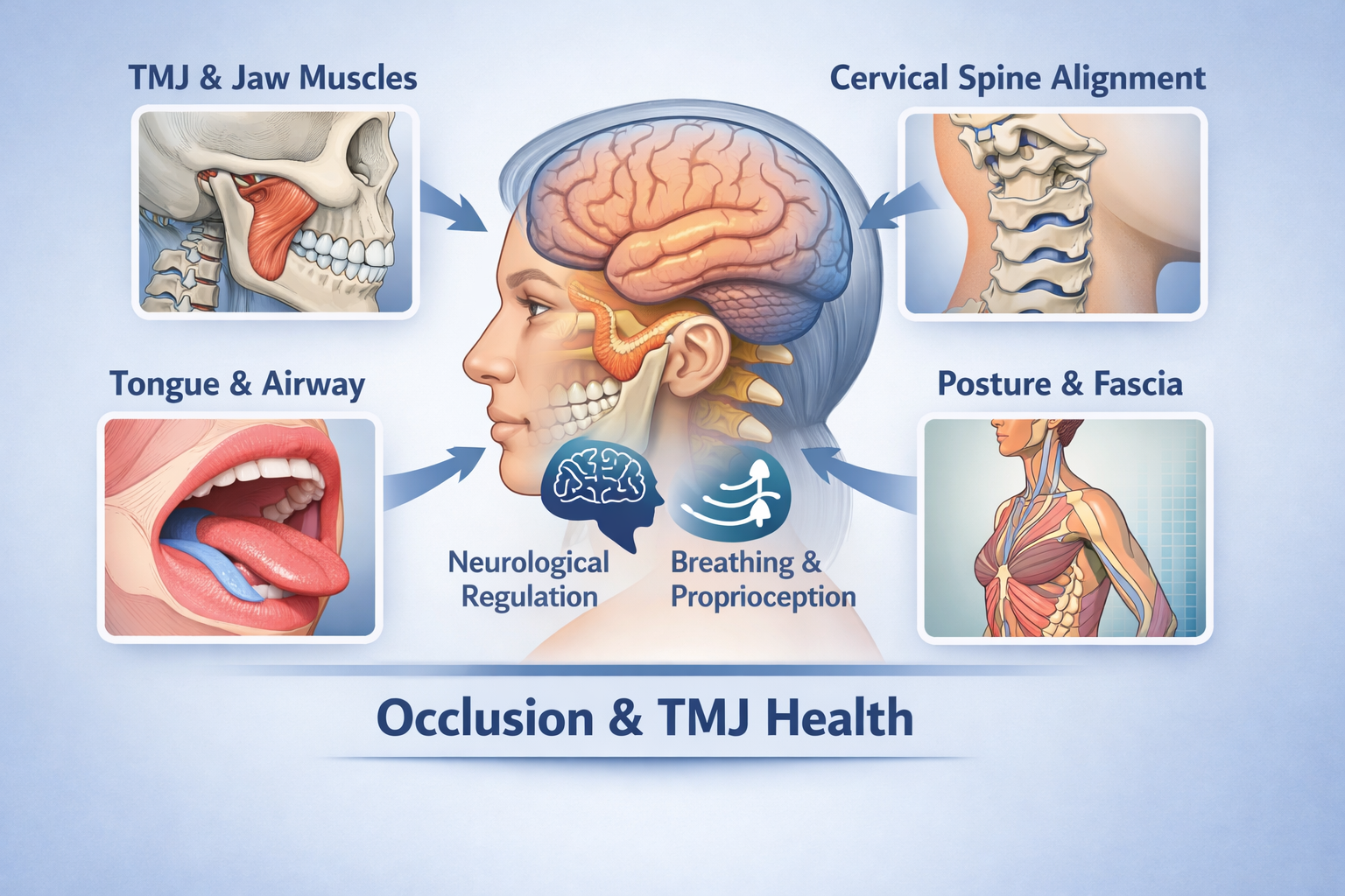
These scenarios highlight an important biological reality: occlusion is not determined by teeth alone, but by how the nervous system, musculoskeletal system, and postural framework support and organize the jaw.
Occlusion Is a Neuro-Musculoskeletal Event, Not Just a Dental One
Mandibular position is governed by a complex interaction between:
The temporomandibular joints
The muscles of mastication
Cervical spine alignment (especially C0–C2)
Proprioceptive input from muscles, ligaments, and joints
Central nervous system regulation
Research in neuromuscular dentistry and orthopedics demonstrates that mandibular position is dynamic, continuously adapting to head posture, cervical alignment, airway demands, and muscle tone rather than existing as a fixed hinge determined solely by tooth contacts.
This explains why a bite can look “perfect” on paper yet feel unstable to the patient: the neuromuscular system is attempting to compensate for imbalance elsewhere. Here is the research on it:
Avivi-Arber & Sessle
PMID: 28853161
https://pubmed.ncbi.nlm.nih.gov/28853161/
The jaw muscles require complex brain processing for precise control during chewing, swallowing, and speaking. This control involves reflexes and coordination between different brain areas that sense jaw position and movement.
Frayne et al.
PMID: 26860522
https://pubmed.ncbi.nlm.nih.gov/26860522/
The lips are better than the jaw at sensing small position changes, even though lip muscles lack spindles (special sensors). This suggests the nervous system uses different methods to sense lip versus jaw position.
Dinsdale et al.
PMID: 32896911
https://pubmed.ncbi.nlm.nih.gov/32896911/
People with TMJ disorders have reduced jaw movement range and weaker bite force. However, there's limited research on whether they can accurately sense jaw position (proprioception).
Miles
PMID: 17257577
https://pubmed.ncbi.nlm.nih.gov/17257577/
When standing still, passive tissue forces hold the jaw in resting position. During running, stretch reflexes in jaw-closing muscles activate automatically to prevent excessive jaw dropping from head movement.
Dinsdale et al.
PMID: 38171215
https://pubmed.ncbi.nlm.nih.gov/38171215/
People with TMJ joint problems have difficulty sensing bite force accurately, especially at lower force levels. Their ability to sense jaw position may also be impaired.
Liu et al.
PMID: 28145597
https://pubmed.ncbi.nlm.nih.gov/28145597/
Sensors in tooth ligaments send signals through the trigeminal nerve to brain areas controlling jaw muscles. Abnormal bite alignment increases these signals, causing jaw muscles to overwork.
Frayne et al.
PMID: 27771297
https://pubmed.ncbi.nlm.nih.gov/27771297/
The right and left sides of the jaw show coordinated position sensing, likely because they work together during chewing. The lips show better position sensing than the jaw joint.
Fujita et al.
PMID: 28194098
https://pubmed.ncbi.nlm.nih.gov/28194098/
When the jaw opens, sensors in jaw-closing muscles activate specific brain regions that process position information. These same brain areas also process pain signals from teeth.
Luo et al.
PMID: 11406816
https://pubmed.ncbi.nlm.nih.gov/11406816/
Muscle spindles in jaw muscles connect to motor control centers through multiple pathways in the brainstem, creating feedback loops that control jaw muscle reflexes and stiffness.
Sato et al.
PMID: 40506626
https://pubmed.ncbi.nlm.nih.gov/40506626/
The cerebellum receives position information from jaw muscle sensors and sends signals back to control jaw movement, mainly through indirect pathways involving multiple relay stations in the brainstem.
TMJ Dysfunction Is Multifactorial by Definition
Temporomandibular disorders (TMD) are widely recognized in the scientific literature as multifactorial conditions, involving:
TMJ structures
Muscles of mastication
Cervical spine biomechanics
Postural control
Psychoneuroendocrine stress responses
Systematic reviews show strong associations between cervical spine dysfunction and TMD symptoms, including pain, restricted range of motion, and altered muscle activity. Interventions addressing cervical posture and muscular imbalance have been shown to reduce TMJ symptoms and improve jaw function—often without any direct dental intervention.
When these contributors are ignored, repeated occlusal adjustments may fail, not because the dentistry is poor, but because the foundation supporting the bite is unstable.
Haller et al.
PMID: 31892357
https://pubmed.ncbi.nlm.nih.gov/31892357/
This analysis of 10 studies found craniosacral therapy significantly reduced chronic pain and improved function compared to usual care, sham treatment, and other manual therapies. Effects lasted up to six months with no serious side effects reported.
Haller et al.
PMID: 26340656
https://pubmed.ncbi.nlm.nih.gov/26340656/
Eight weekly craniosacral therapy sessions significantly reduced chronic neck pain compared to fake treatment. Pain relief, improved function, and better quality of life lasted for three months after treatment ended.
Muñoz-Gómez et al.
PMID: 35160211
https://pubmed.ncbi.nlm.nih.gov/35160211/
A four-week craniosacral therapy program reduced migraine pain intensity, how often migraines occurred, disability, and medication use compared to fake treatment. Benefits remained at two-month follow-up in migraine patients.
Castro-Sánchez et al.
PMID: 27347698
https://pubmed.ncbi.nlm.nih.gov/27347698/
Ten craniosacral therapy sessions improved pain intensity, oxygen levels in blood, blood pressure, and blood mineral levels more than massage in chronic low back pain patients, with effects lasting one month.
Castro-Sánchez et al.
PMID: 20702514
https://pubmed.ncbi.nlm.nih.gov/20702514/
Twenty weeks of craniosacral therapy reduced pain at 13 of 18 tender points in fibromyalgia patients and improved heart function measurements. Pain reduction lasted one year after treatment.
Matarán-Peñarrocha et al.
PMID: 19729492
https://pubmed.ncbi.nlm.nih.gov/19729492/
Twenty-five weeks of craniosacral therapy improved anxiety, sleep quality, and quality of life in fibromyalgia patients compared to fake treatment. Most benefits lasted six months, while sleep improvements lasted one year.
Haller et al.
PMID: 33647398
https://pubmed.ncbi.nlm.nih.gov/33647398/
In primary care, craniosacral therapy (average 7 sessions) significantly improved symptoms, function, and quality of life across 114 different health conditions in children and adults with minimal side effects.
Jäkel & von Hauenschild
PMID: 23131379
https://pubmed.ncbi.nlm.nih.gov/23131379/
This review of seven studies found craniosacral therapy showed positive results for pain reduction and well-being, but study quality was moderate, indicating more rigorous research is needed.
Downey et al.
PMID: 17154138
https://pubmed.ncbi.nlm.nih.gov/17154138/
In rabbits, forces similar to those used in craniosacral therapy did not move skull bones or change brain pressure, suggesting craniosacral therapy works through different mechanisms than moving bones.
Upper Cervical Alignment and Mandibular Position
The upper cervical spine—particularly the occiput, atlas (C1), and axis (C2)—plays a critical role in:
Head posture
Trigeminal nerve signaling
Muscle recruitment patterns
Proprioceptive input affecting jaw position
Altered alignment or mobility in this region can change mandibular resting position and functional movement patterns. Studies have shown correlations between forward head posture, altered cervical curvature, and changes in mandibular kinematics and muscle activation.
From a clinical standpoint, this means:
A patient may occlude differently depending on head posture
The jaw may function in a compensated position to protect cervical structures
Occlusal contacts may shift as cervical tone normalizes
Upper cervical chiropractic care aims to reduce abnormal joint stress and restore more symmetrical neuromuscular signaling—allowing the mandible to function from a more balanced reference point. Here is the research on it:
Xiao et al.
PMID: 36776487
https://pubmed.ncbi.nlm.nih.gov/36776487/
TMD patients with jaw joint pain show more forward head posture than those without joint pain. The worse the TMD symptoms, the more pronounced the forward head position, especially in men.
Yu et al.
PMID: 40447683
https://pubmed.ncbi.nlm.nih.gov/40447683/
Combining jaw exercises with neck exercises improves mouth opening and reduces jaw muscle stiffness better than neck exercises alone in people with abnormal neck posture from smartphone use.
Oleksy et al.
PMID: 34660797
https://pubmed.ncbi.nlm.nih.gov/34660797/
Treating only the neck in people with neck pain improved their jaw function, even though they didn't complain of jaw pain initially, showing the neck and jaw are connected.
Minervini et al.
PMID: 37048735
https://pubmed.ncbi.nlm.nih.gov/37048735/
This analysis of multiple studies confirms a connection between body posture and TMJ disorders, likely through shared nerve pathways that control both jaw and body position.
Cuenca-Martínez et al.
PMID: 32872670
https://pubmed.ncbi.nlm.nih.gov/32872670/
People with TMJ disorders have more neck disability and reduced neck movement, plus lower pain thresholds in neck muscles, but their neck alignment isn't necessarily different from healthy people.
Opris et al.
PMID: 36078792
https://pubmed.ncbi.nlm.nih.gov/36078792/
X-ray measurements of neck posture and a small bone in the throat may help understand TMJ disorders, and conservative treatments targeting these areas might reduce symptoms.
Yao et al.
PMID: 36241923
https://pubmed.ncbi.nlm.nih.gov/36241923/
Forward head posture itself doesn't directly cause jaw muscle pain in TMD patients, but neck muscle pain is strongly linked to jaw muscle pain.
Hong et al.
PMID: 30554035
https://pubmed.ncbi.nlm.nih.gov/30554035/
In elderly people with TMJ and neck pain, spine degeneration (especially in upper neck bones) relates to forward head posture and painful trigger points in jaw and neck muscles.
Ferreira et al.
PMID: 31292981
https://pubmed.ncbi.nlm.nih.gov/31292981/
Women with TMJ disorders have limited upper neck movement and weak deep neck muscles. These problems correlate with both neck disability and jaw pain levels.
Silveira et al.
PMID: 25883963
https://pubmed.ncbi.nlm.nih.gov/25883963/
Jaw problems and neck problems are highly correlated. People with more tender muscles in the upper back and temples have worse jaw and neck function.
Rocha et al.
PMID: 24118029
https://pubmed.ncbi.nlm.nih.gov/24118029/
This review found the relationship between head/neck posture and TMJ disorders is still unclear due to poor quality studies, so more research is needed.
La Touche et al.
PMID: 32181811
https://pubmed.ncbi.nlm.nih.gov/32181811/
Manual therapy and exercises targeting the neck reduce TMJ pain and increase pain tolerance in jaw muscles, showing neck treatment helps jaw problems.
Calixtre et al.
PMID: 30307636
https://pubmed.ncbi.nlm.nih.gov/30307636/
Upper neck mobilization and neck strengthening exercises significantly reduced facial pain and headaches in women with TMJ disorders compared to no treatment.
Neuromuscular, Fascial, and Airway Interactions
The TMJ does not function in isolation. It is embedded within a continuous myofascial and neurological network extending from the cranium through the cervical spine and into the thorax and pelvis.
Tongue Posture and Restrictions
Restricted tongue mobility (including ankyloglossia) can alter:
Swallowing mechanics
Mandibular movement patterns
Cervical muscle tone
Airway dynamics
Compensatory muscle activity may increase strain on the TMJs and alter occlusal perception over time. This is well-documented in airway-focused orthodontics and orofacial myology literature. Here are some studies on it:
Cai et al.
PMID: 26796752
Account required
Sign in to see more
Create an account or sign in to unlock the full list.